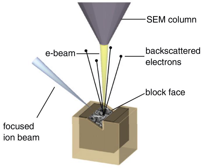
The EMCF has implemented a Focused Ion Beam-Scanning Electron Microscope (FIB-SEM, see figure above), together with the Schwab team and in collaboration with Carl Zeiss Microscopy. This imaging technology relies on combining serial etching of a resin block with the scanning of the exposed surface. When the cell features (membranes, cytoskeletal elements, protein complexes) are cross-sectioned, they are revealed in an almost equivalent way as with a TEM microscope. The sequential slicing and viewing steps therefore lead to the acquisition of large volumes in an automated way.
The main advantage compared to serial TEM or serial tomography is the size of the volume that can be acquired; with a close to one thousand-fold increase in favor to the FIB-SEM.
The fact that this technique allows imaging sub-cellular features at high resolution and in 3D opens to the study of membrane trafficking, organelle architecture and cytoskeletal arrangement in the volume of the cell, but also cell-cell or cell-matrix interactions. The goal is to make this new microscope a versatile tool for several projects. The EMCF strives to become a specialized center for the application of such approaches and will actively contribute to the methodological development necessary to make it a key technique in biology.
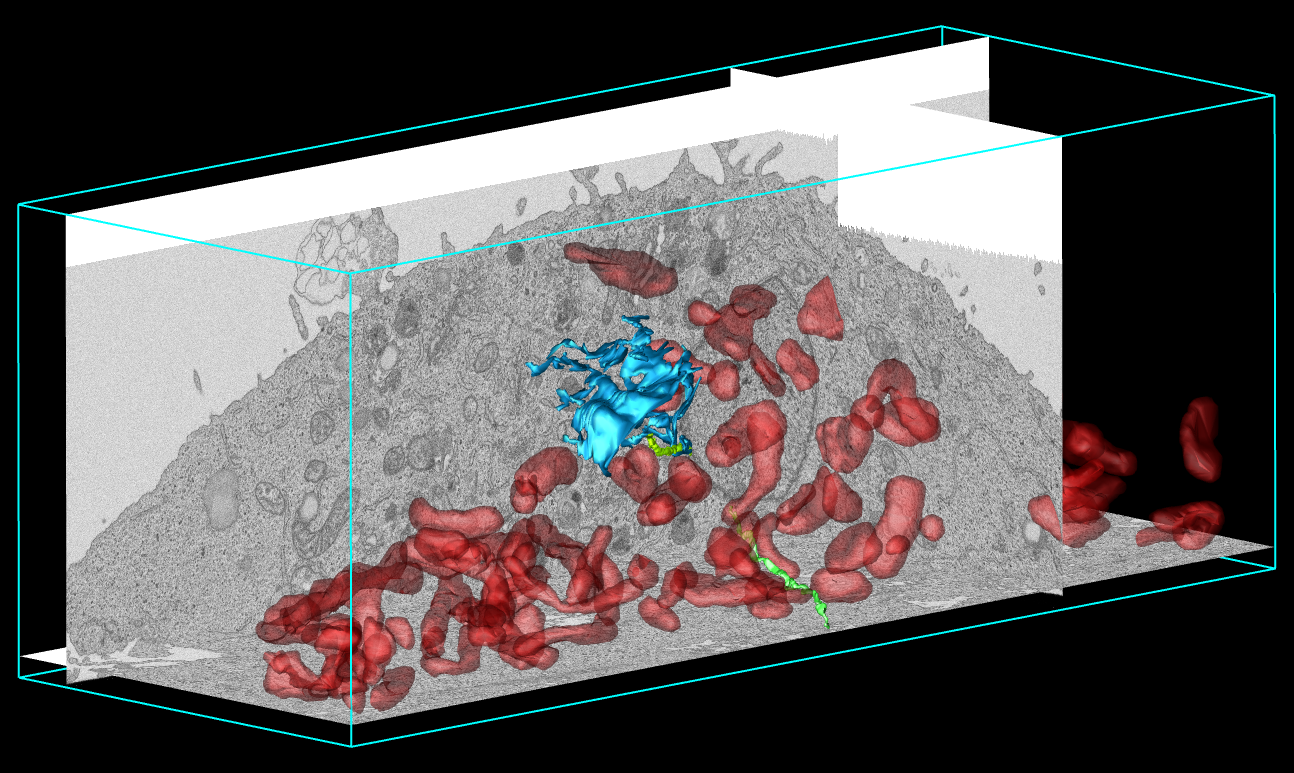
3D datasets such as the one shown that was acquired acquired using the FIB-SEM from a cultured cell, allow an understanding of the volume organization within cells. Mitochondrias are segmented in red, a fraction of the ER (in blue) shows interactions with the plasma membrane specializations (green). (courtesy of Anna Steyer, Schwab team)