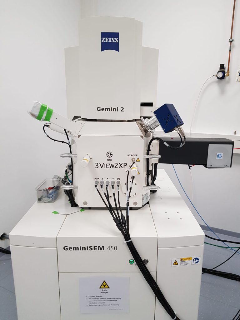
The Serial Block Face – Scanning Electron Microscope (SBF-SEM or SBEM) is an automated instrument that allows 3D electron microscopy of large samples over days or weeks. It combines iterative imaging with a SEM and physical slicing of the sample with a diamond knife mounted on a microtome inside the SEM chamber. In the EMCF we have a Gatan 3view system mounted on a Zeiss Gemini 450 SEM. The x,y resolution that can be achieved is similar to that of a FIB-SEM (~5nm pixel size), but the z resolution is more limited (~30nm).
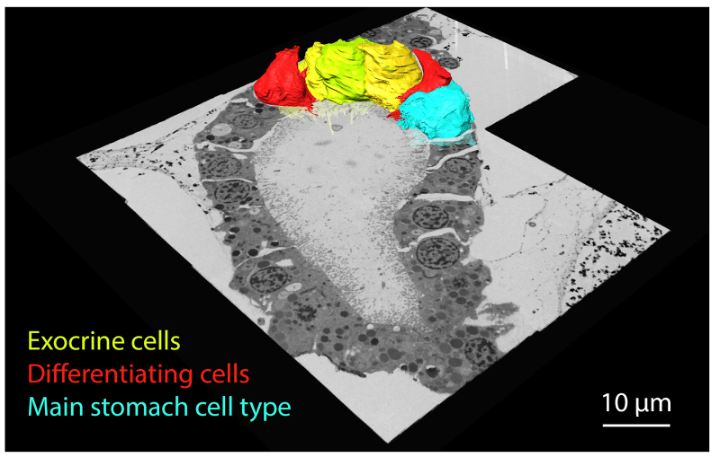
This instrument is typically used to image large tissues and small organisms up to several hundred microns in all dimensions (e.g. tissues or full Platynereis dumerilii larvae, sea urchin larvae, brain slices, mosquito gut), when high isotropic resolution is not necessary. Examples of samples recently acquired in the EMCF are mouse tissues (brain), sea urchin larvae, Platynereis dumerilii tissues, mosquito tissues, human leukocytes.