For specimens that do not permit targeting using Light Microscopy, all available information is obtained using EM.
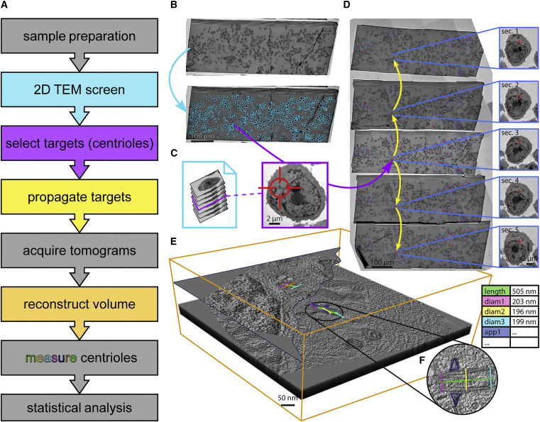
In order to efficiently find rare or sparsely spread phenotypes, we employ a TEM screening approach. We use automated acquisition using SerialEM driven by image analysis that identifies target features (like cells) in a low-magnification overview of the specimen. At the target features, we can then acquire high-resolution TEM data in 2D or 3D tomography to unveil the underlying ultrastructure.
Software tools for automated transmission electron microscopy.
Schorb M, et al. Nat Methods. 2019 Jun;16(6):471-477.
This approach was also used to characterise centrioles in cancer cells:
Dittrich T and Köhrer S, et al., Multi-Modality Imaging Reveals Structural Centrosome Aberrations As a Potential Driver of Chromosomal Instability in Early-Stage Plasma Cell Disorders. Blood. 2021 November 23; 138;1. https://doi.org/10.1182/blood-2021-150747
As well as in moss spermatids:
Gomes Pereira S, et al. The 3D architecture and molecular foundations of de novo centriole assembly via bicentrioles. Current Biology. 2021 Oct 11;31(19):4340-4353.e7. (https://doi.org/10.1016/j.cub.2021.07.063).