Room temperature ultramicrotomes:
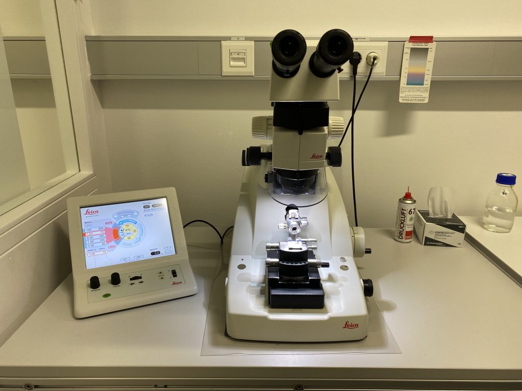
Resin sections are indispensable for the observation of biological material with a TEM, as samples have to be thinned in order to become electron-transparent. Plastic sections are made using such ultramicrotomes operated at room temperature. The EMCF is equipped with 2x UC7 ultramicrotomes (Leica Microsystems, ). We use diamond knives attached to a water trough to collect and manipulate the sections. As the sections are cut, they glide off of the knife and are floated onto the surface of the water. The sections are collected as ribbons and can be picked up by carbon-coated formvar grids or grids coated purely with carbon.
Cryo-ultramicrotome:
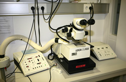
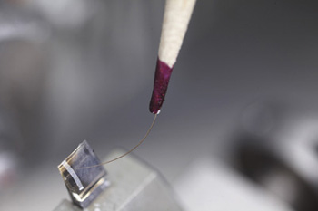
The EMCF has one cryo-ultramicrotome dedicated for Tokuyasu cryosectioning, a technique that is widely used for immuno-labelling purposes. Samples are trimmed at -90oC into a geometrical shape, and then sectioning occurs at around ~ -110oC. Sections are guided onto the diamond knife by an eyelash tool and then picked up with a loop of methylcellulose and placed onto an EM grid.