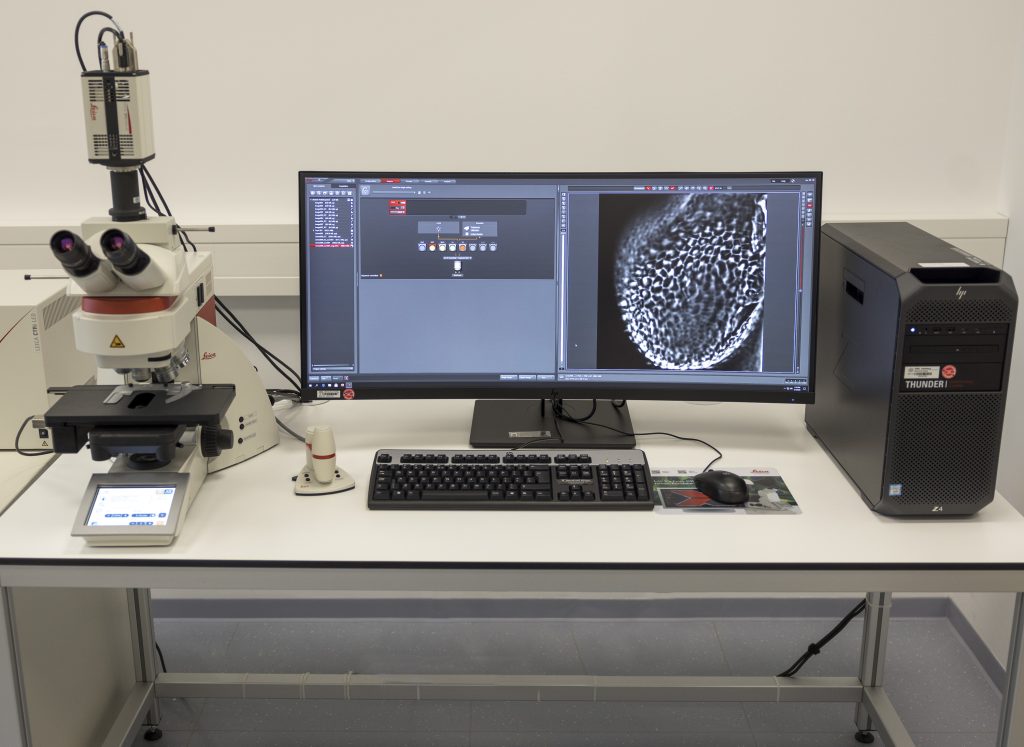
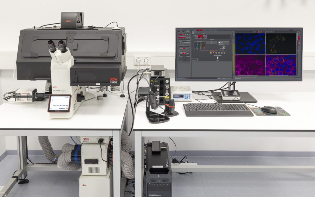
THUNDER Imager Live Cell
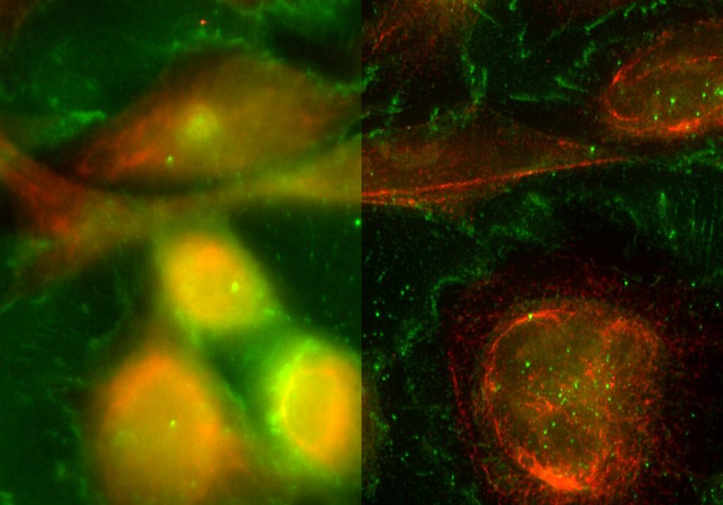
THUNDER is an opto-digital technology from Leica Microsystems that uses the Computational Clearing method for haze removal to generate high-resolution and high-contrast images.
A THUNDER Imager Live Cell provides a complete microscopy imaging system for minimally invasive and precise live-cell imaging. High-speed and/or long term examination of specimens, e.g. cell cultures and organoids, can be performed at low phototoxicity and photobleaching.
Features
- sCMOS camera technology
- Efficient removal of out-of-focus blur in real time with Computational Clearing
- Automation of the imaging and analysis workflow with the LAS X software
- Processing and reconstruction using the Aivia software platform
Specifications
- Based on the fully motorised, high-end, inverted research microscope DMi8
- High-speed positioning with the Quantum stage and Synapse real-time controller
- High-speed illumination with a multi-line LED light source
- Fast switching external filter wheel
- Adaptive Focus Control (AFC) with closed loop focus
- Climate chamber ensures optimal physiological conditions for living cells
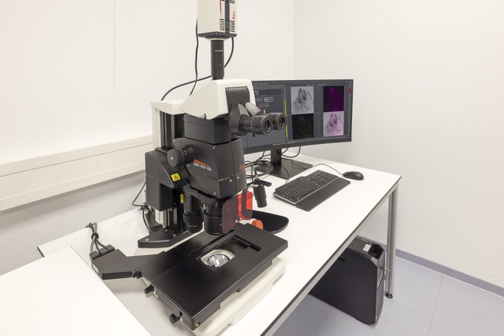
THUNDER Imager Model Organism
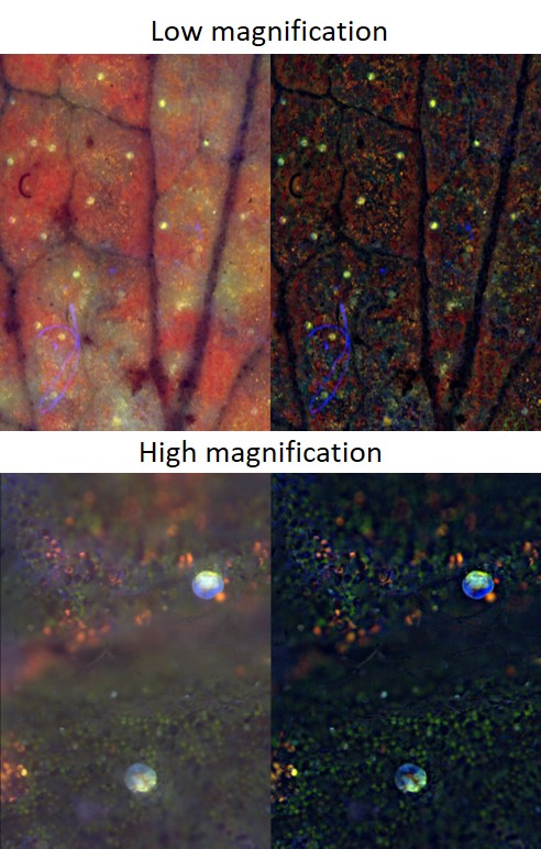
The THUNDER Imager Model Organism from Leica Microsystems allows fast and easy 3D exploration of whole organisms for developmental or molecular biology research using Computational Clearing.
It can be used for studying, e.g., Drosophila, C. elegans, zebrafish, plants, and mice. Model organisms can be studied from overview to the details.
Features
- Rapid acquisition of blur-free images showing fine details, even from deep within thick organisms
- Large model organisms
- Imaging and analysis workflow within the LAS X operation software
Specifications
- Based on Leica M205 FCA stereo microscope
- LMT scanning stage and motor focus drive
- Equipped with a high-end sCMOS camera
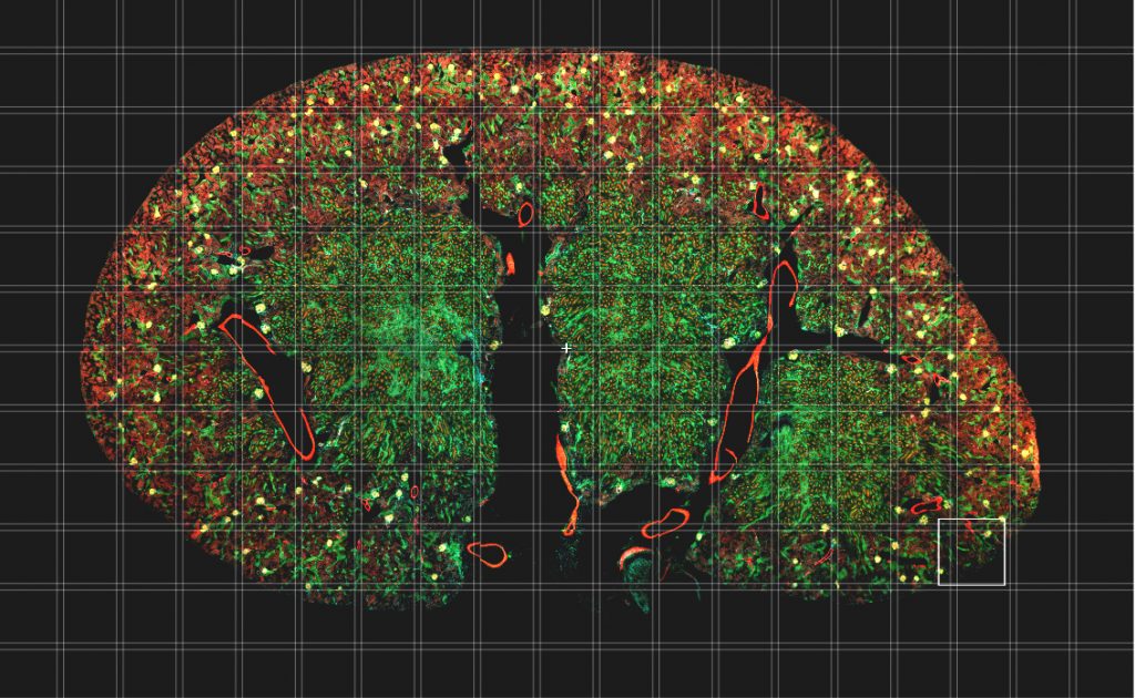
THUNDER Imager 3D Tissue
The THUNDER Imager Tissue from Leica Microsystems allows real-time fluorescence imaging of 3D tissue sections typically used for neuroscience and histology research. It also enables the acquisition of rich, detailed images of thick tissues. These images are free of haze from out-of-focus blur because of Computational Clearing for haze removal. Structures like axons and dendrites of neurons in a brain slice can be imaged.
Features
- Rapidly acquisition of blur-free images even deep within thick sections
- Get fast overviews of whole tissue sections
- Imaging and analysis workflow within the LAS X operation software
- Processing and reconstruction of complex images with the Aivia software platform
Specifications
- Based on a fully automated upright research microscope for the acquisition of multi-color 3D images
- sCMOS camera system
- Software creates blur-free large overviews of the entire tissue specimen
- Precise motorised z-focus drive to capture images in the z-direction and visualise them with the 3D Viewer