Important note: This system will be available in open access after the opening of the EMBL IC.
With the first commercially available MINFLUX from Abberior Instruments, the EMBL IC enables scientists to image a broad range of biological samples (living/fixed) with a spatial resolution of < 5 nm. This allows to study among others the structure of protein complexes like nuclear pores with unprecedented 3D resolution. Additionally, MINFLUX tracking allows to study movement of single molecules in living cells with up to 10 kHz sampling rate (one localisation each 100 µs, which is up to 100 x faster than conventional camera-based tracking).
Features of our MINFLUX system:
- MINFLUX imaging with 1-3 nm localisation precision in 2D/3D
- Tracking of single molecules with up to 10 kHz featuring <20 nm resolution
- Nanometre-level active sample stabilization system based on fiducial markers in the sample
Specifications of our MINFLUX microscope:
- Laser lines: MINFLUX: 640 nm, 561 nm, Confocal: 488 nm, 405 nm
- Two-colour ratiometric MINFLUX imaging with 640 nm or 560 nm excitation
- IX83 inverted microscope stand equipped with a 1.4 NA 100x oil objective
News:
Abberior and the European Molecular Biology Laboratory (EMBL) have launched a collaboration on the application of MINFLUX microscopy in biological research. The goal of this collaboration is to combine forces regarding further development of the method, and to apply superresolution MINFLUX microscopy to living systems (see details).
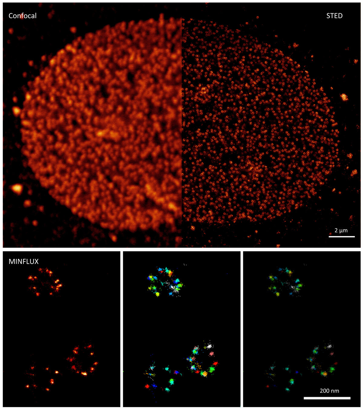
(Credit: Sebastian Schnorrenberg/EMBL Imaging Centre)