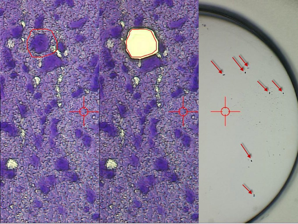
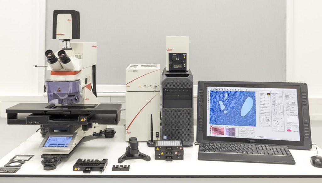
LMD7
The LMD7 laser microdissection microscope from Leica Microsystems enables users to isolate specific microscopic target areas under visual control for downstream molecular biology analysis. Regions of interest (ROI) can be isolated from entire areas of tissue, like cell clusters or tumours, down to single cells or even subcellular structures, such as chromosomes. Live cell dissection for downstream analysis or re-cultivation (cloning) as well as laser manipulation is possible. The LMD7 is equipped with a LMT350 scanning stage which allows collection of dissectates directly into regular PCR tubes or multi-well plates, such as 96-well PCR plates. Downstream analysis is typically used for genomics (DNA), transcriptomics (mRNA, miRNA) by qPCR, Microarray, RNA-seq, next generation sequencing (NGS), proteomics, metabolomics done with Western blots, and mass spectrometry. Lipidomics can be done with laser microdissection.
Features
- Laser movement by optics
- Direct collection into 96-well plates simply by gravity
Specifications
- Wavelength: 349 nm
- Pulse frequency: 10–5,000 Hz
- Pulse length: <4 ns
- Average pulse energy: 120 μJ
- Range of dedicated LMD objectives: 2.5x, 5x, 10x, 20x, 40x, 63x, and 150x
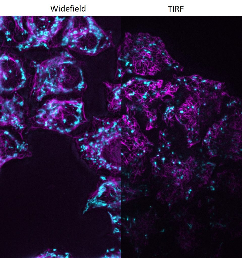
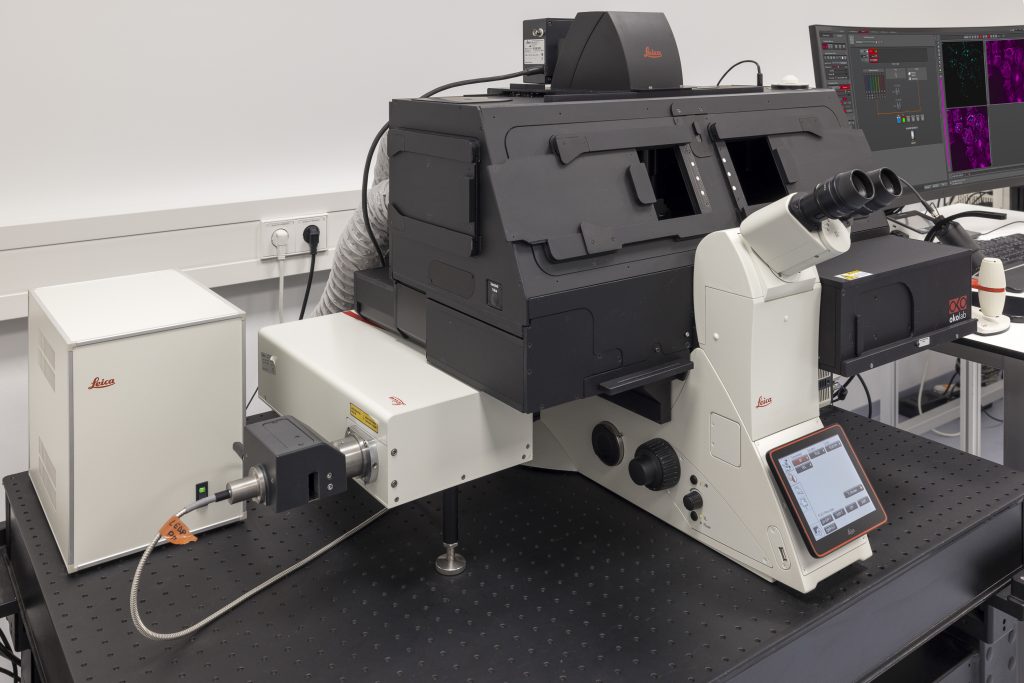
DMi8 S with scanner and TIRF module
DMi8 S from Leica Microsystems is a live cell imaging system to execute advanced experiments with imaging modalities like photomanipulation or TIRF, both simultaneously or sequential. For dynamic processes at the cell surface TIRF (Total Internal Reflection Fluorescence) microscopy is the method of choice to visualize single molecules with super-resolution by maximizing the fluorescent signal- to-noise-ratio. Furthermore, the system can be set-up for multispectral photomanipulation techniques like FRAP, acceptor bleaching, activation, optogenetics, DNA damage, or ablation within one single experiment. Imaging specialists are on site to support your particular application.
Features
- Infinity TIRF module with azimuth shifting capability to optimise the illumination field.
- Infinity scanner for advanced multispectral photomanipulation applications (enables to bleach, cut, activate, stimulate).
- LAS X Navigator software to create quick overviews of the sample and identify the important details instantly. High resolution image acquisition can be set up using templates for slides, dishes, and multi-well plates.
Specifications
- Fully motorized DMi8 inverted research microscope with integrated real-time controller to operate the system with microsecond precision
- Adaptive Focus Control (AFC)
- Equipped with a high-end sCMOS camera