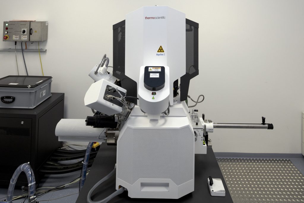
Edit

EMBL Imaging Centre

Open access to cutting-edge electron and light microscopy

We provide researchers from Europe and beyond with a synergistic portfolio of imaging services including cryo-EM, super-resolution and intravital microscopy to enable new ground-breaking research that crosses the scales of biology.

Cryo-FIB Milling for Lamella Preparation

Crossbeam 550

The Crossbeam 550 (focused ion beam-scanning electron microscope) from ZEISS allows data acquisition in 3 dimensions at isotropic nanometer voxel size. Samples from cells to tissues and organisms can be looked at with electron microscopic detail. Chemically fixed, high-pressure frozen and freeze-substituted as well as purely frozen samples can be analyzed. On top of data production, the Crossbeam550 is also equipped to generate cryo-lamellae to be analyzed further in a transmission electron microscope e.g. using tomography.

Unique features

  • Isotropic volume acquisition down to a few nm
  • Large 3D volume acquisition
  • Cryo lamella preparation

Important specifications

  • Detectors: InLens, EsB and Se
  • Quorum Cryo-SEM preparation system
  • Zeiss Atlas 5 software
High-pressure frozen and freeze substituted HeLa cell imaged at 5 nm voxel size with the Crossbeam550.
Credit: Stuart Ingham/EMBL.
Zeiss Crossbeam 550. Credit: Stuart Ingham/EMBL.

Aquilos 2

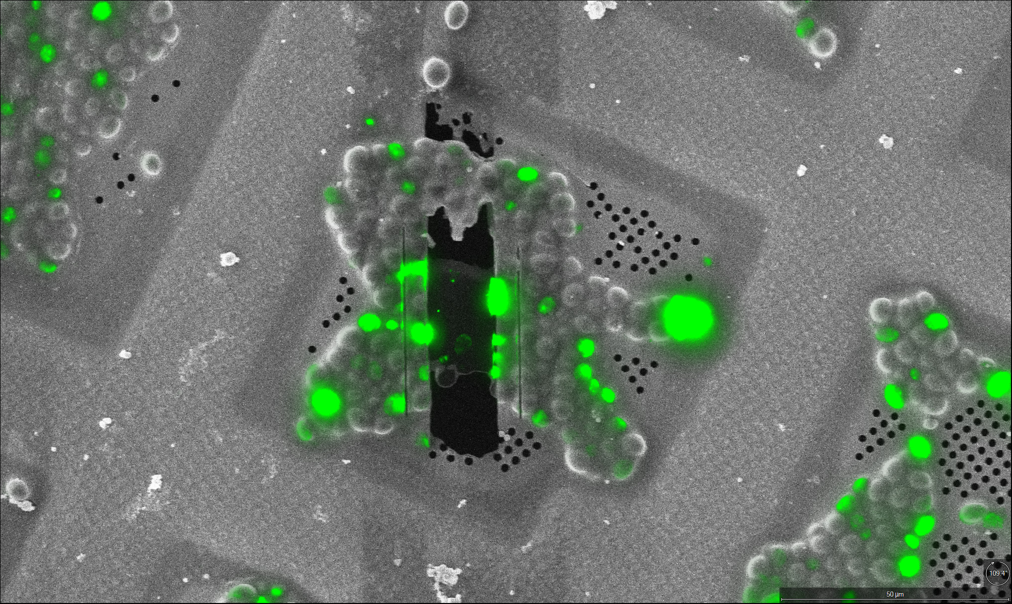
The Aquilos 2 (focused ion beam – scanning electron microscope) from Thermo Fisher Scientific enables the preparation of cellular lamellae from vitreous samples suitable for high-resolution cryo-electron tomography analyses. Plunge frozen as well as high-pressure frozen samples can be processed at cryo temperature by standard FIB milling and cryo-lift out approaches, respectively. Automated FIB milling routines based on commercial and in-house developed software packages enable unsupervised overnight operations for high-throughput milling.

Unique features

  • Automated cryo lamella preparation
  • Nanomanipulator for cryo-lift out
  • Integrated widefield fluorescence microscopy

Important specifications

  • Serial-FIB and AutoTEM software for automated lamella preparation
  • EasyLift NanoManipulator
  • Integrated Fluorescence Light Microscope (iFLM)
Lamella through plunge frozen yeast cells imaged with the iFLM inside the Aquilos 2.
Thermo Fisher Scientific Aquilos 2. Credit: Stuart Ingham/EMBL.
Edit