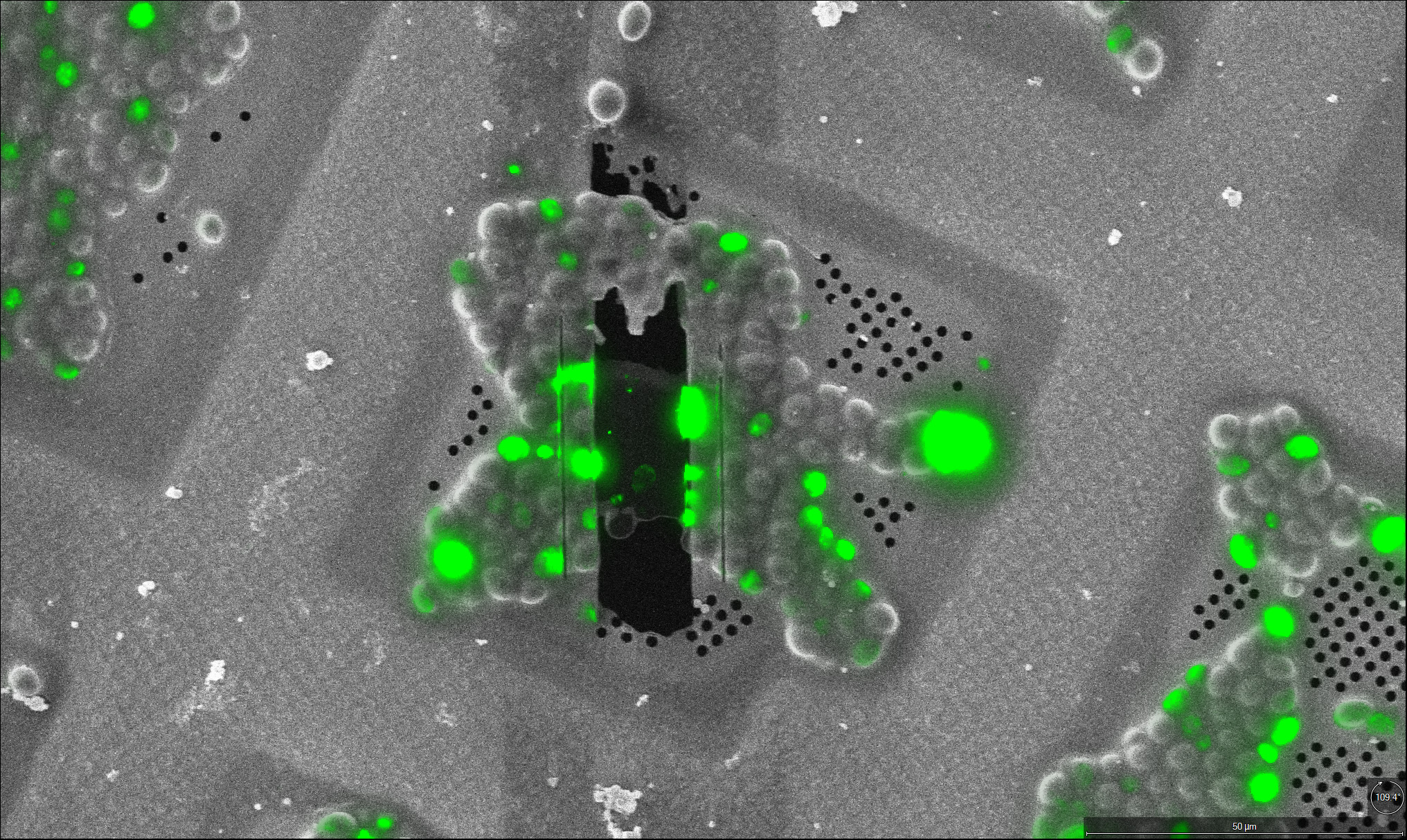
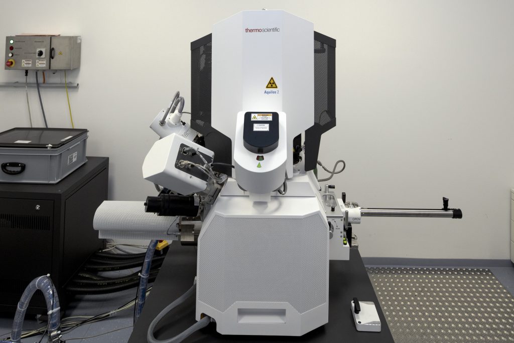
Aquilos 2
The Aquilos 2 (focused ion beam – scanning electron microscope) from Thermo Fisher Scientific enables the preparation of cellular lamellae from vitreous samples suitable for high-resolution cryo-electron tomography analyses. Plunge frozen as well as high-pressure frozen (HPF) samples can be processed at cryo temperature by standard FIB milling and cryo-lift out approaches, respectively. The lamellae production and lift out process can be guided with fluorescence signal, either performed on a cryo-light microscope prior to the session, or with integrated fluorescence microscope in Aquilos 2.Automated FIB milling routines based on commercial and in-house developed software packages enable unsupervised overnight operations for high-throughput milling.
Important specifications
- Serial-FIB and AutoTEM software for automated lamella preparation
- EasyLift NanoManipulator
- Integrated Fluorescence Light Microscope (iFLM)
Offered workflows
- Automated over-night lamella preparation on plunge frozen samples.
- Waffle milling on HPF waffle grids.
- Serial lift out on HPF waffle or planchette carriers, followed with automated lamellae prepraration.
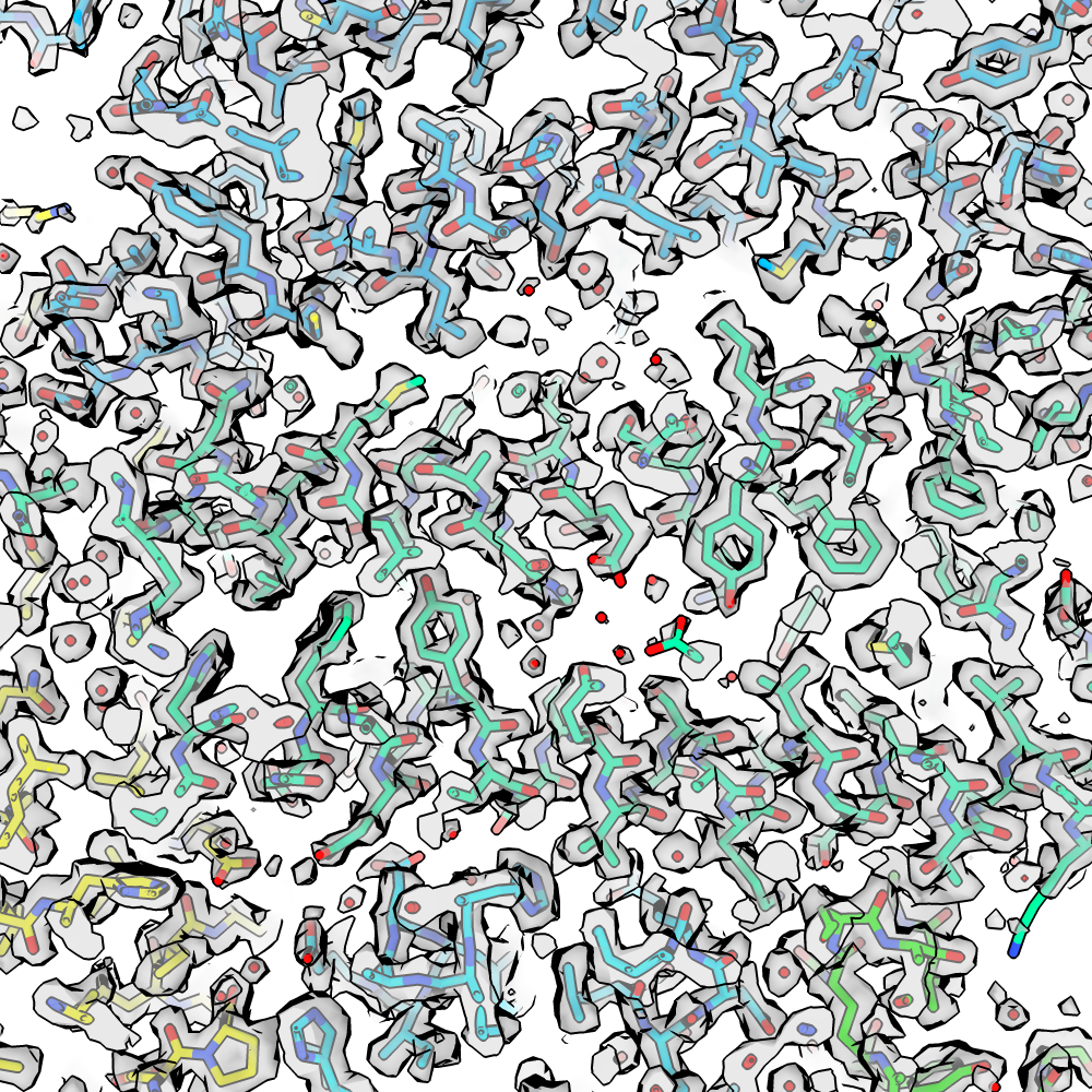
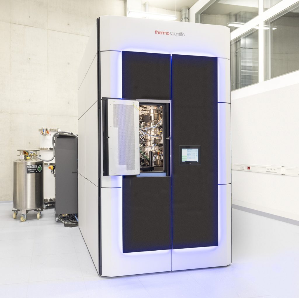
Titan Krios G4
The Titan Krios G4 from Thermo Fisher Scientific is a 300 kV transmission electron microscope (TEM) with a three-condenser lens system. It features the latest technology for high-end single-particle and tomography data acquisition including a cold field emission gun (C-FEG), SelectrisX energy filter, Falcon 4 electron counting camera and volta phase plate. It’s configured for high-throughput automated single particle and tomography data acquisition.
Unique features
- C-FEG electron source with narrow energy spread
- State-of-the-art energy filter and electron counting camera
- phase plate
- Autoloader system for easy sample transfer between TFS TEMs
Important specifications
- 300 kV C-FEG electron source
- SelectrisX energy filter
- Falcon 4EC direct electron detector
- Volta phase plate
- SerialEM software for tailored, automated data acquisition