Elizabeth Duke
Lead
EMBL Hamburg
Next generation imaging for biology across scales
IMAGINE will provide technologies to probe structure and function of biological specimens in their natural context
At the core of the IMAGINE project is the development of innovative cross-scale imaging instrumentation, tools, and methods, which will advance the state-of-art of the participating pan-European RIs and show their transformative operational potential for the Life Sciences to address major socio-economic challenges of human and planetary health.
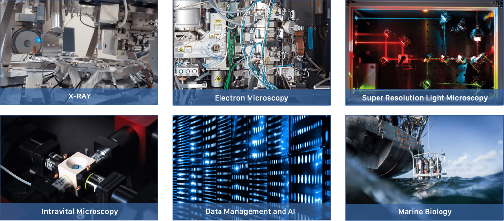
In six work packages (WPs) IMAGINE will develop instrumentation, tools and methods related to X-ray imaging (WP1), cryo-EM (WP2), super-resolution microscopy (WP3) and intravital imaging (WP4), image-based sample screening and preservation in the field (WP5). Hereby, a specific focus lies on the correlation and integrated use of all of the technologies. IMAGINE aims at making those imaging technologies sufficiently robust for service provision and allow their integration into cross-scale experiments both in terms of the specimen handling and the AI-powered data analysis and integration (WP6).
This work package focusses on using hard X-ray imaging of biological samples and aims at developing sample handling techniques and instrumentation to enable routine experiments at both room and cryogenic temperatures. A workflow will be generated for non-specialist users and deployed at different European RI’s and used for correlative imaging. The WP will explore the potential of X-ray imaging of biological matter in a near native state.
The work package is co-led by EMBL Hamburg and ESRF. Additional partners include EMBL Grenoble, Paul Scherrer Institute and ARINAX.
Significant progress was made in developing X-ray imaging workflows, including a high-throughput room-temperature tomography pipeline and high-resolution cryo-tomography methods. New sample preparation protocols, silicon nitride supports, and cross-compatible mounting systems enable automated, multimodal, and cross-facility imaging workflows. The approaches were successfully demonstrated on diverse biological, physiological, and environmental samples, support large-scale studies, and are being integrated with electron and light microscopy for advanced multimodal analysis.
WP1 advanced high-throughput and cryogenic tomography workflows, improving automated 3D imaging of diverse biological samples. Cross-platform sample mounting systems were developed, and protocols for correlative X-ray/electron tomography were refined. The HiTT pipeline was successfully demonstrated on sponges, Drosophila brains, and plankton, with deep learning reconstructions enabling imaging of dose-sensitive specimens, validating the pipeline’s versatility for multi-scale structural analysis.
This project is funded by the European Union (GA#101094250). Views and opinions expressed are however those of the authors only and do not necessarily reflect those of the European Union. Neither the European Union nor the granting authority can be held responsible for them.

This work package focusses on electron microscopy and aims at developing new sample preparation methods, sample carriers, and integrative workflows for seamless transfer and correlative imaging between different modalities, including X-ray tomography (WP1), volume imaging, cryo-ET, and advanced LM (WP3) approaches.
The work package is co-led by EMBL Heidelberg and Max Planck Institute of Biochemistry. Additional partners include EMBL Grenoble, Arinax, Rosalind Franklin Institute, TESCAN, Leiden University Medical Centre, Leica, and ESRF.
Period 1
WP2 team developed and tested advanced workflows for automated cryo-sample preparation and multimodal cellular imaging. The EasyGrid platform enables rapid, high-quality vitrification of adherent and suspension cells, improving preservation over traditional methods. New silicon nitride sample carriers support cross-modality workflows combining X-ray nano-imaging, cryo-FIB milling, and cryo-ET. Integrated fluorescence and FIB-SEM systems enable precise targeting, while high-throughput plasma milling accelerates lamella production from multicellular specimens. Initial experiments show improved structural preservation and feasibility for advanced multimodal imaging workflows, with further proof-of-concept studies planned for 2024.
Period 2
WP2 has developed integrated workflows for high-quality cryo-imaging of single cells and multicellular specimens. Automated jet vitrification using the EasyGrid platform improves preservation of thick cellular regions, while new SiN supports and modified Autogrids enable compatibility with both hard X-ray tomography and cryo-ET. High-pressure freezing protocols have been optimized for diverse multicellular samples, and the integration of cryo-fluorescence microscopy into FIB-SEM instruments allows precise 3D targeting of regions of interest, including deeply buried structures. Serial Lift-Out methods and plasma-FIB milling using argon and xenon ions have increased throughput, reliability, and automation for lamella production, supporting large-scale, reproducible cryo-ET studies. These advances together establish scalable, standardized pipelines for correlative multi-modal imaging, bridging X-ray, FIB-SEM, and cryo-ET, and provide a robust foundation for broad user access and community adoption.
This project is funded by the European Union (GA#101094250). Views and opinions expressed are however those of the authors only and do not necessarily reflect those of the European Union. Neither the European Union nor the granting authority can be held responsible for them.
WP3 aims to establish super-resolution fluorescence microscopy to bridging physiological function to molecular structure determination. It will develop both dynamic and cryo-SRM in order to image both structure and real-time function of molecular machines in living cells. WP3 works closely with WP4 and WP2 on correlative intravital imaging and cryo-EM, and WP5 on environmental samples.
The work package is led by EMBL Heidelberg. Additional partners include KTH Royal Institute of Technology, Leiden University Medical Centre, and Leica Microsystem.
Period 1
Significant progress has been made in developing advanced cryo-super-resolution and MINFLUX imaging workflows. Fluorophores have been systematically characterised at room and cryogenic temperatures for photostability, quantum yield, and reversible switching to enable new correlative imaging approaches. Automated software tools were developed for tomogram segmentation and fluorescence-guided particle picking, supporting precise correlation of cryo-SR and Cryo-EM data. Robust procedures for screening and labelling cells on EM grids, including in vivo dyes, genetic tags, and antibody staining, have been established to facilitate multiplexed super-resolution imaging.
Period 2
WP3 develops dynamic MINFLUX imaging of molecular machines and approaches for cryo-super-resolution microscopy. Self-blinking dyes enable repeated imaging, while dual-color MINFLUX tracks conformational changes with nanometer precision. Instrument upgrades and optimized workflows, including cryo-SMLM and automated software for particle picking, allow precise correlative targeting in cryo-CLEM. Screens of small fluorophores identified reversible cryo-dark states for super-resolution and workflows for high-precision, dynamic, and multi-modal imaging of molecular structures are being developed.
This project is funded by the European Union (GA#101094250). Views and opinions expressed are however those of the authors only and do not necessarily reflect those of the European Union. Neither the European Union nor the granting authority can be held responsible for them.
This WP targets intravital imaging and its correlative use with complementary imaging technologies in WP1-3. It will develop a multi-modal volume-imaging platform to study cellular and tissue dynamics in mesoscopic and freely-moving specimens and advance correlative imaging strategies and workflows of non-transparent tissues and organisms to link dynamics to underlying (ultra-)structure. Close synergies exist with WP5 on deployment in the field, and WP6 on image restoration, real-time analysis and integration.
The work package is led by EMBL Heidelberg. Additional partners include EMBL Barcelona, EMBL Rome, Human Technopole, Centro de Ciências do Mar Algarve, and Paul Scherrer Institute.
Period 1
Significant progress has been made in developing high-speed, multi-modal imaging platforms for living organisms. Improvements to the mesoscopic oblique plane microscope enhanced axial resolution and enabled imaging of freely behaving marine organisms. In parallel, the integration of microfluidics for high-throughput studies has been started. The label-free optical coherence microscope was miniaturized for mobile deployments, maintaining high-resolution imaging of various marine species. Correlative intravital imaging workflows are being established for non-transparent tissues such as Drosophila, combining fluorescence, X-ray tomography, and electron microscopy, supported by multi-modal probe development.
Period 2
WP4 advanced multi-modal intravital imaging of living and non-transparent organisms by integrating Optical Coherence Microscopy (OCM) and Oblique Plane Microscopy (OPM) into a single platform, enabling high-speed, volumetric, label-free and fluorescence imaging of freely behaving specimens. The mobile OCM and optimized OPM with active tracking were further successfully tested on diverse marine and laboratory models. In parallel, cross-modal approaches were developed to link functional imaging of Drosophila sLNv neurons with structural data via X-ray tomography (ESRF) and EM, enabling future correlative imaging workflows. Finally, a library of ∼450 dyes with high X-ray contrast was established to support future correlative labeling. Together, these efforts yielded dynamic, high-resolution imaging across scales, bridging function, structure, and behavior in near-native conditions.
This project is funded by the European Union (GA#101094250). Views and opinions expressed are however those of the authors only and do not necessarily reflect those of the European Union. Neither the European Union nor the granting authority can be held responsible for them.
This work package aims at developing field platforms for high-resolution/high-throughput phenotypic imaging-based species identification coupled to automated sorting at different size scales for subsequent analyses by the novel cross-scale imaging technologies developed in WP1-4. The phenotyping will rely on AI-based image data analysis provided by WP6 and will enable for the first time acquisition of combined morphological/taxometric and molecular/ultrastructural features in single specimens collected in their environmental niches.
The work package is led by EMBL Heidelberg. Additional partners include Sorbonne University, University of Vigo, and Centro de Ciências do Mar Algarve.
Period 1
WP5 team has advanced high-speed, image-enabled workflows for sorting marine microorganisms. Automated microscopy and fluorescence-based labeling allow identification and isolation of specific plankton phenotypes, while high-throughput imaging generates large annotated datasets for AI model development. Mesoscopic imaging using a compact OCM has been successfully deployed in the field to visualize larger marine organisms, laying the groundwork for image-based sorting. These technologies have been demonstrated and taught in training courses, with further AI-driven sorting development planned.
Period 2
WP5 advances image-enabled sorting of marine organisms across scales, from single cells to mesoscopic plankton. Task 5.1 established high-speed flow cytometry workflows for unicellular plankton (<50 μm), linking high-resolution microscopy to sorter-acquired images, enabling expert- and AI-guided gating strategies with >90% predictive accuracy for target taxa. Task 5.2 extended these approaches to larger organisms (50 μm–2 mm) by integrating Optical Coherence Microscopy (OCM) and Oblique Plane Microscopy (OPM) with custom 3D-printed microfluidic chips and high-frame-rate imaging, laying the groundwork for automated, real-time sorting. Together, these developments create field-deployable platforms for isolating and analyzing ecologically important plankton, supporting downstream multi-scale imaging, omics, and ecological studies.
This project is funded by the European Union (GA#101094250). Views and opinions expressed are however those of the authors only and do not necessarily reflect those of the European Union. Neither the European Union nor the granting authority can be held responsible for them.
WP6 aims at developing novel image analysis solutions to allow integration of the multi-scale data output from the different technologies developed in WP1-5 and their correlative analysis. It will be driven by AI-based tools for image restoration and segmentation as well as by representation learning for microscopic and mesoscopic objects of biological interest.
The work package is co-led by EMBL Heidelberg and EMBL-EBI. Additional partners include Human Technopole and Centro de Ciências do Mar Algarve.
Period 1
Progress has been made in AI-based analysis and computational tools for advanced microscopy. Algorithms have been developed for morphological screening, multi-view imaging, cross-modality super-resolution, and image decomposition to enable high-quality reconstruction and reduced color usage in fluorescence imaging. Early work on segmentation and 3D registration of cells and subcellular structures supports correlative light, X-ray, and electron microscopy. Additionally, cloud-ready multimodal data management and visualization solutions using OME-NGFF formats have been prototyped and tested.
Period 2
WP6 advanced AI-driven image analysis for cross-scale biological imaging, developing 3D and 2D shape descriptors for sorting, photon-efficient denoising and structure separation (µSplit → MicroSplit), and automated 3D registration for correlating light and electron microscopy. A standardized multimodal data model with RO-Crate enables consistent dataset integration and cloud-based visualization. These tools support high-throughput morphology-based sorting, noise mitigation, and cross-scale analysis across IMAGINE workflows.
This project is funded by the European Union (GA#101094250). Views and opinions expressed are however those of the authors only and do not necessarily reflect those of the European Union. Neither the European Union nor the granting authority can be held responsible for them.