
Niki Keklikoglou
HCMR
Next generation imaging for biology across scales
IMAGINE will provide technologies to probe structure and function of biological specimens in their natural context

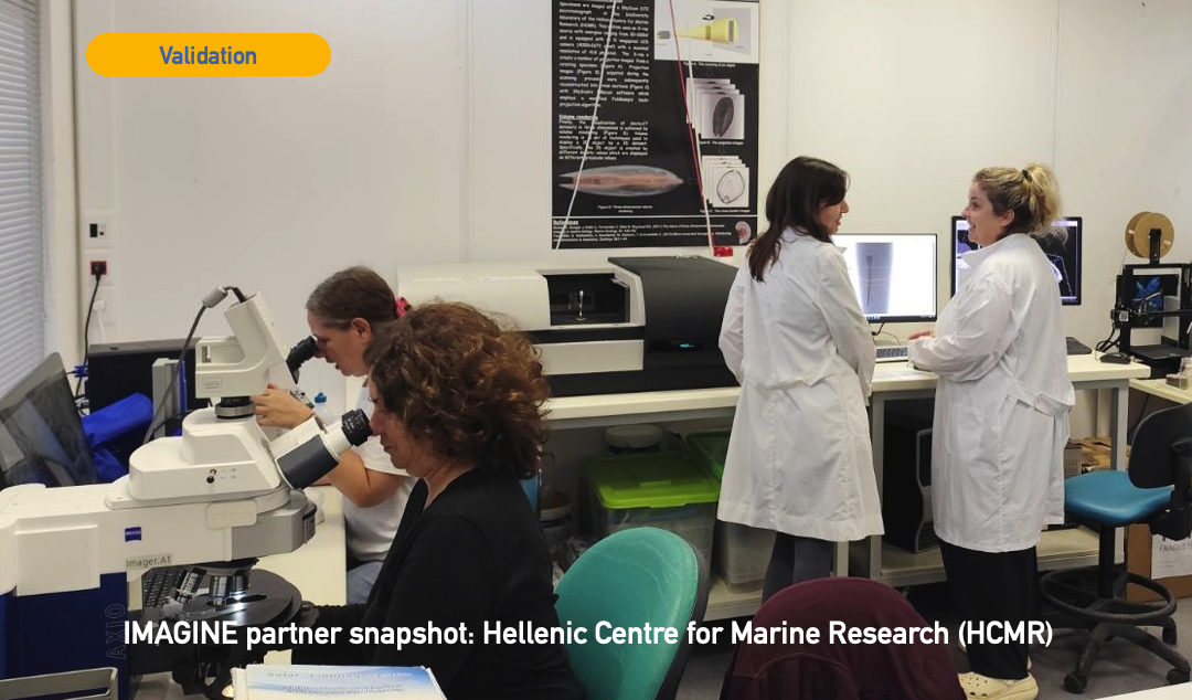
Marine organisms are at the front line of climate change, but studying how they respond often means cutting, staining or drying precious samples. Within IMAGINE, the Hellenic Centre for Marine Research (HCMR) is helping to show that there is a better way.
HCMR is a partner in Work Package 9 (WP9), led by EMBRC-ERIC, which tests whether the imaging technologies developed in IMAGINE are ready to serve marine researchers. HCMR’s first test case, with the X-ray imaging technology of WP1, is a sponge.
HCMR brings marine biology expertise and real research questions. EMBL Hamburg and the IMAGINE X-ray team bring cutting-edge imaging. EMBRC-ERIC connects the two, so that new technologies are shaped by the needs of marine researchers from the start.
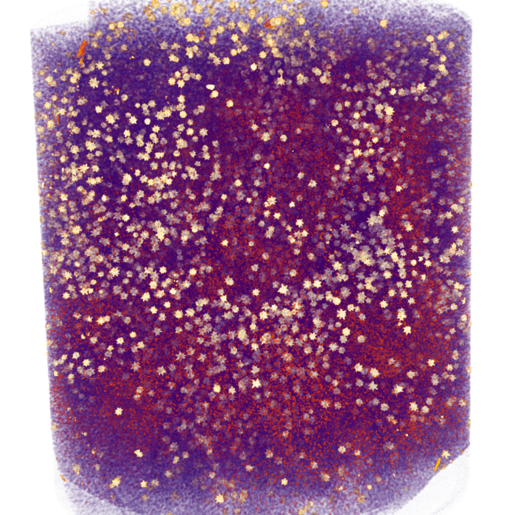
Sponges and other benthic invertebrates that live attached to the seabed cannot move away when conditions around them change, making them particularly vulnerable to environmental pressures. HCMR studies how climate change, including ocean acidification and rising temperature, affects the form and structure of these animals. Within the framework of the national MACCIMO project, one of the species in focus is the sponge Chondrilla nucula, sampled from different populations and from experimental treatments simulating future ocean conditions.
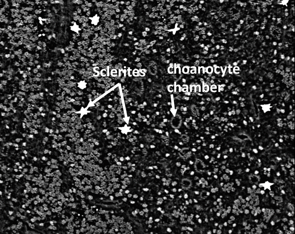
HCMR had already scanned these sponges with a conventional micro-CT (SkyScan 1172). The images gave a good overview, but the resolution was too low to show fine internal structures such as sclerites, the microscopic skeletal elements. The samples also needed extra preparation: they were dried with hexamethyldisilazane (HMDS) to improve tissue contrast. This is where IMAGINE’s new X-ray technology comes in.
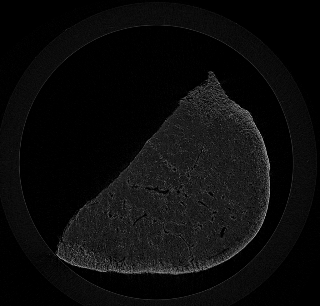
26 specimens were sent to EMBL Hamburg for phase-contrast high-throughput X-ray tomography (HiTT) at the EMBL P14 beamline on the PETRA III synchrotron at DESY. This technology can reach cellular-level resolution, using a voxel size of 660nm³ at high acquisition speeds. The samples were fixed in 4% formalin, dehydrated and stored in 70% ethanol. Because HiTT has a field of view of ~1mm x ~1mm works on each specimen was trimmed into two pieces of about 1 mm³, one from the exterior and one from the interior of the sponge, so both regions could be compared.
Ksenia Denisova (EMBL Hamburg) collected and analysed the HiTT data and generated the images that made it possible to investigate the sponge specimens at cellular-level resolution. Her work provided the essential imaging data for the cross-scale analysis in IMAGINE, enabling comparisons between the internal and external regions of the sponges and helping to bridge structural observations across different scales
Beyond this one sponge, the technology gives researchers better tools to track how climate change affects marine biodiversity. Rare and irreplaceable specimens, such as natural history museum collections, could be digitised without damage. And because the method is fast, more samples can be studied in less time.
Niki’s Quote: “Advances in imaging and the combination of different imaging techniques can reveal how marine organisms are structured and how they function, helping us understand how they respond to changing environmental conditions.”

This project is funded by the European Union (GA#101094250). Views and opinions expressed are however those of the authors only and do not necessarily reflect those of the European Union. Neither the European Union nor the granting authority can be held responsible for them.