Anna Akhmanova
University of Utrecht, The Netherlands
EMBO | EMBL Symposium
EMBO and EMBL are committed to sharing research advances and sustaining scientific interaction throughout the coronavirus pandemic. We are delighted to announce that this conference is going virtual and invite you to join us online. The virtual conference includes talks from invited speakers, short talk presenters, digital poster sessions, online group discussions and networking opportunities.
Access to the platform: please note we have sent several email invitations to join platform, if you haven’t received it, please check your spam folder. If it’s not there, please contact Iva Gavran (iva.gavran@embl.de).
For late registrations without submission please use this link.
Got something to say? Tweet it! #EESImaging
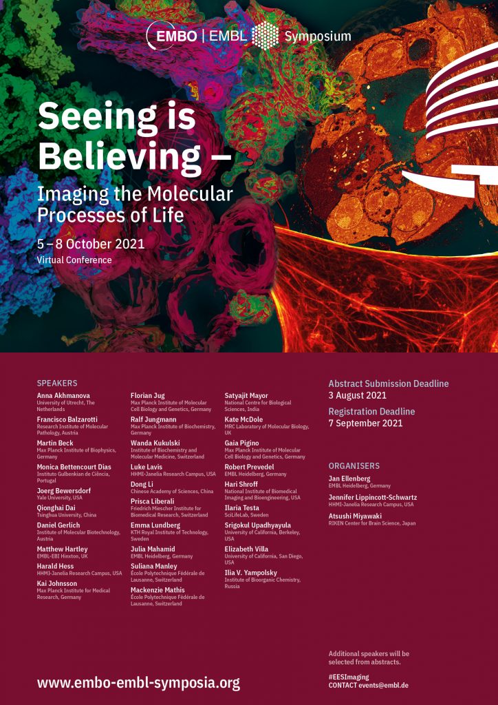
The molecular processes of life are naturally dynamic in space and time from the atomic to the organismal level. The rapid development of imaging methods across the full scale of biological organisation is revolutionising our ability to visualise the inner workings of macromolecular complexes, organelles, cells, tissues, organs and whole organisms. Being able to see biological processes unfold in real time allows us to understand the mechanisms of life as well as disease.
The symposium will bring together the leading developers of imaging methods with cutting-edge applications that illustrate how imaging can answer biological questions. We will emphasize methods that can capture the dynamics of life, spanning the whole range from molecular resolution to imaging of whole organisms.
From its beginning in 2011, Seeing is Believing has embraced novel imaging technologies that open new windows for biological discovery, including single-molecule and super-resolution, light sheet and correlative light electron microscopy.
In this spirit, in 2021, we will include a session on in situ structural biology, prominently featuring 3D cryo-electron microscopy. Recent exciting examples show that these technologies have the potential to resolve the structures of molecular machines in action in their natural cellular and even organismal context, seamlessly linking it to other scales of imaging in the symposium. Everybody interested in the latest imaging technologies and their applications in the life sciences should attend.
The symposium provides many opportunities for presentations, discussions, and interactions between students, postdocs, junior, and senior investigators.
“A great event to immerse yourself in the newest ideas and approaches in bio-imaging.” Andreas Gahlmann, University of Virginia, USA
“It’s a fantastic opportunity to find great scientists sharing new insights on technological and biological advancement in a vibrant environment.” Simone Bonaccorsi, UNSW Sydney, Australia
“This remains my favourite conference, with the blend of technology and applied sciences, I challenge any scientist not to come out of this meeting feeling exhilarated and full of ideas.” Dr. Ola Sabet, University of Zürich, Switzerland
“I learned an exceptional amount about the state of the art in super resolution microscopy and its applications by attending this conference, which would have otherwise taken months to sift through the literature to fine the relevant papers. I’m definitely planning on coming back for the next edition.” Greg McMahon, Principal Research Scientist, National Physical Laboratory Teddington, UK
“Seeing is Believing is a fantastic meeting that brings together the exciting, rapidly developing fields of cutting-edge microscopy development and cell biology. Please please please keep organising these meetings, they are very inspirational for young scientists and exceptionally well-received.” Kobus van Unen, Cell Biology Institute, University of Bern, Switzerland
University of Utrecht, The Netherlands
Research Institute of Molecular Pathology
Austria

Max Planck Institute of Biophysics
Germany
Instituto Gulbenkian de Ciência
Portugal
Yale University
USA

Tsinghua University
China
Institute of Molecular Biotechnology
Austria
EMBL EBI Hinxton
UK

HHMI-Janelia Research Campus
USA

Max Planck Institute for Medical Research
Germany

Fondazione Human Technopole
Italy
Max Planck Institute of Biochemistry
Germany
University of Bern
Switzerland

HHMI-Janelia Research Campus
USA

Chinese Academy of Sciences
China
Friedrich Miescher Institute for Biomedical Research
Switzerland
KTH Royal Institute of Technology
Sweden
EMBL Heidelberg
Germany
École Polytechnique Fédérale de Lausanne
Switzerland
École Polytechnique Fédérale de Lausanne
Switzerland
National Centre for Biological Sciences
India

MRC Laboratory of Molecular Biology
UK

Max Planck Institute of Molecular Cell Biology and Genetics
Germany

EMBL Heidelberg
Germany
National Institute of Biomedical Imaging and Bioengineering
USA
SciLifeLab
Sweden

University of California, Berkeley
USA

EMBL Heidelberg
Germany
HHMI-Janelia Research Campus
USA
RIKEN Center for Brain Science
Japan
EMBL Heidelberg
Germany
EMBL Heidelberg
Germany
All times in the programme below are shown as the time in Europe/Berlin.
To find out the equivalent time zone in your location, enter Berlin, the programme time and your city into the Time Zone Converter.
| Time (Europe/Berlin) | Speaker |
|---|---|
| 14:00 – 14:50 | Pre-symposium webinars:
|
| 15:00 – 15:50 | Pre-symposium webinars:
|
| 16:00 – 16:50 | Pre-symposium webinars:
|
| 17:00 – 17:50 | Pre-symposium webinars:
|
| Time (Europe/Berlin) | Speaker |
|---|---|
| 14:00 – 14:10 | Opening remarks (Livestreamed and available on demand in Video Library until 22 Oct 2021) |
| 14:10 – 16:20 | Session 1: Super-resolved imaging |
| 14:10 – 14:15 | Session intro Chair: Suliana Manley (Livestreamed and available on demand in Video Library until 22 Oct 2021) |
| 14:15 – 14:30 | Invited lecture: Adaptive temporal sampling with an intelligent iSIM Suliana Manley – École Polytechnique Fédérale de Lausanne, Switzerland (Lecture will be livestreamed and available on demand in Video Library until 22 Oct 2021) |
| 14:30 – 14:45 | Invited lecture: Live cell compatible super resolution fluorescence microscopy for 3D imaging Ilaria Testa – SciLifeLab, Sweden (Lecture will be livestreamed and available on demand in Video Library until 22 Oct 2021) |
| 14:45 – 15:00 | Invited lecture: Accessing the nano-scale with MINFLUX Francisco Balzarotti – Research Institute of Molecular Pathology, Austria (Lecture will be livestreamed and available on demand in Video Library until 22 Oct 2021) |
| 15:00 – 15:15 | Invited lecture: All-optical super-resolution imaging of molecules in their nanoscale cellular context Joerg Bewersdorf – Yale University, USA (Lecture will be livestreamed and available on demand in Video Library until 22 Oct 2021) |
| 15:20 – 15:30 | Short break |
| 15:30 – 15:45 | Molecular organization and mechanics of vimentin single filaments revealed by SMLM Cecile Leduc – Institute Jacques Monod, France (Lecture will be livestreamed and available on demand in Video Library until 22 Oct 2021) |
| 15:45 – 16:00 | Podosome cluster dynamics unveiled by radial spatiotemporal image correlation spectroscopy and super-resolution imaging Paul Wiseman – McGill University, Canada (Lecture will be livestreamed and available on demand in Video Library until 22 Oct 2021) |
| 16:00 – 16:15 | Visualizing the native cellular organization by coupling cryo-fixation with expansion microscopy (Cryo-ExM) Marine Laporte – University of Geneva, Switzerland (Lecture will be livestreamed and available on demand in Video Library until 22 Oct 2021) |
| 16:20 – 16:50 | Meet the speakers of Session 1 Suliana Manley, Ilaria Testa, Francisco Balzarotti, Joerg Bewersdorf, Cecile Leduc, Paul Wiseman, Marine Laporte |
| 16:50 – 18:05 | Poster session 1 Topic 1: Super-resolved imaging Topic 2: Correlative imaging Topic 7: Discoveries by bioimaging (presenters with last name A-F) |
| 18:05 – 20:15 | Session 2: Correlative imaging |
| 18:05 – 18:10 | Session intro Chair: Harald Hess (Livestreamed and available on demand in Video Library until 22 Oct 2021) |
| 18:10 – 18:25 | Invited lecture: 3D Electron Microscopy and Cryo-CLEM Harald Hess – HHMI-Janelia Research Campus, USA (Lecture will be livestreamed and available on demand in Video Library until 22 Oct 2021) |
| 18:25 – 18:40 | Invited lecture: Multimodality Structured illumination microscopy for super-resolution live-cell imaging Dong Li – Chinese Academy of Sciences, China (Lecture will be livestreamed and available on demand in Video Library until 22 Oct 2021) |
| 18:40 – 18:55 | Invited lecture: Visualising the architecture of organelle contact sites Wanda Kukulski – University of Bern, Switzerland (Lecture will be livestreamed and available on demand in Video Library until 22 Oct 2021) |
| 18:55 – 19:10 | Invited lecture: Revealing mitotic chromosome assembly by light- and electron microscopy Daniel Gerlich – Institute of Molecular Biotechnology, Austria (Lecture will be livestreamed and available on demand in Video Library until 22 Oct 2021) |
| 19:15 – 19:25 | Short break |
| 19:25 – 19:40 | Super-resolution and correlative microscopy reveal atypical centrosome organization of the malaria-causing parasite Plasmodium falciparum Caroline Simon – Heidelberg University Hospital, Germany (Lecture will be livestreamed and available on demand in Video Library until 22 Oct 2021) |
| 19:40 – 19:55 | Measuring conformational changes in clathrin light chain at single sites of endocytosis with FLIM-FRET-CLEM Kazuki Obashi – NHLBI, NIH, USA (Lecture will be livestreamed and available on demand in Video Library until 22 Oct 2021) |
| 19:55 – 20:10 | Correlative light and electron microscopy suggests that mutant huntingtin dysregulates the endolysosomal pathway in presymptomatic Huntington’s disease Ya Zhou – University College London, UK (Lecture will be livestreamed and available on demand in Video Library until 22 Oct 2021) |
| 20:15 – 20:45 | Meet the speakers of Session 2 Harald Hess, Dong Li, Wanda Kukulski, Daniel Gerlich, Caroline Simon, Kazuki Obashi, Ya Zhou |
| 20:45 – 20:50 | Short break |
| 20:50 – 21:35 | Social programme: speed networking |
| Time (Europe/Berlin) | Speaker |
|---|---|
| 13:15 – 13:45 | Industry session |
| 14:00 – 16:10 | Session 3: In situ structural biology |
| 14:00 – 14:05 | Session intro Chair: Julia Mahamid (Livestreamed and available on demand in Video Library until 22 Oct 2021) |
| 14:05 – 14:20 | Invited lecture: Molecular views into cellular functions by in-cell cryo-electron tomography Julia Mahamid – EMBL Heidelberg, Germany (Lecture will be livestreamed and available on demand in Video Library until 22 Oct 2021) |
| 14:20 – 14:35 | Invited lecture: Towards a mechanistic understanding of motile and primary cilia with CLEM and cryo-electron tomography Gaia Pigino – Max Planck Institute of Molecular Cell Biology and Genetics, Germany (Lecture will be livestreamed and available on demand in Video Library until 22 Oct 2021) |
| 14:35 – 14:50 | Invited lecture: Opening Windows into the Cell: Bringing structure to cell biology using Cryo-electron tomography Elizabeth Villa – University of California, San Diego, USA (Lecture will be livestreamed and available on demand in Video Library until 22 Oct 2021) |
| 14:50 – 15:05 | Invited lecture: Conformational dynamics of nuclear pore complexes Martin Beck – Max Planck Institute of Biophysics, Germany (Lecture will be livestreamed and available on demand in Video Library until 22 Oct 2021) |
| 15:10 – 15:20 | Short break |
| 15:20 – 15:35 | Enterovirus cell entry visualized in situ by cryo-electron microscopy Pavel Plevka – CEITEC, Masaryk University, Czech Republic (Lecture will be livestreamed and available on demand in Video Library until 22 Oct 2021) |
| 15:35 – 15:50 | In-cell structural biology of actin filament nucleation by the Arp2/3 complex studied by cryo-electron tomography and subtomogram averaging Florian Schur – Institute of Science and Technology (IST) Austria, Austria (Lecture will be livestreamed and available on demand in Video Library until 22 Oct 2021) |
| 15:50 – 16:05 | Motion of single molecular tethers reveals dynamic subdomains at organelle contact sites Christopher Obara – HHMI – Janelia Research Campus, USA (Lecture will be livestreamed and available on demand in Video Library until 22 Oct 2021) |
| 16:10 – 16:40 | Meet the speakers of Session 3 Julia Mahamid, Gaia Pigino, Elizabeth Villa , Martin Beck , Pavel Plevka, Florian Schur, Christopher Obara |
| 16:40 – 17:55 | Poster session 2 Topic 3: In situ structural biology Topic 4: Image analysis Topic 7: Discoveries by bioimaging (presenters with the last name G-S) |
| 17:55 – 20:05 | Session 4: Image analysis |
| 17:55 – 18:00 | Session intro Chair: Florian Jug (Livestreamed and available on demand in Video Library until 22 Oct 2021) |
| 18:00 – 18:15 | Invited lecture: Microscopy Image Restoration and Downstream Analysis – recent improvements and hopes for the future Florian Jug – Fondazione Human Technopole, Italy (Lecture will be livestreamed and available on demand in Video Library until 22 Oct 2021) |
| 18:15 – 18:30 | Invited lecture: Using machine learning for measuring movement Mackenzie Mathis – École Polytechnique Fédérale de Lausanne, Switzerland (Lecture will be livestreamed only) |
| 18:30- 18:45 | Invited lecture: Image-based spatiotemporal dissection of the human proteome Emma Lundberg – KTH Royal Institute of Technology, Sweden (Lecture will be livestreamed and available on demand in Video Library until 22 Oct 2021) |
| 18:45 – 19:00 | Invited lecture: Multiscale intravital fluorescence microscopy Qionghai Dai – Tsinghua University, China (Lecture will be livestreamed and available on demand in Video Library until 22 Oct 2021) |
| 19:05 -19:15 | Short break |
| 19:15 – 19:30 | Invited lecture: The BioImage Archive: Enabling image data publication, sharing and reuse Matthew Hartley – EMBL-EBI Hinxton, UK (Lecture will be livestreamed and available on demand in Video Library until 22 Oct 2021) |
| 19:30 – 19:45 | Learning a representation of cellular morphology: Unsupervised deep learning for shape and texture characterization of a cell Valentyna Zinchenko – EMBL Heidelberg, Germany (Lecture will be livestreamed and available on demand in Video Library until 22 Oct 2021) |
| 19:45 – 20:00 | An interactive deep learning-based approach reveals the organelle ultrastructure Yusuke Hirabayashi – The University of Tokyo, Japan (Lecture will be livestreamed and available on demand in Video Library until 22 Oct 2021) |
| 20:05 – 20:35 | Meet the speakers of Session 4 Florian Jug, Mackenzie Mathis, Emma Lundberg, Qionghai Dai, Matthew Hartley, Valentyna Zinchenko, Yusuke Hirabayashi |
| 20:35 – 20:40 | Short break |
| 20:40 – 21:40 | Social programme: quiz icebreaker and bar mixer |
| Time (Europe/Berlin) | Speaker |
|---|---|
| 13:15 – 13:45 | Industry session |
| 14:00 – 16:10 | Session 5: Organismal imaging |
| 14:00 – 14:05 | Session intro Chair: Kate McDole |
| 14:05 – 14:20 | Invited lecture: Imaging mammalian development with light-sheet microscopy Kate McDole – MRC Laboratory of Molecular Biology, UK (Lecture will be livestreamed only) |
| 14:20 – 14:35 | Invited lecture: New optical tools to interrogate biological function in-vivo Robert Prevedel – EMBL Heidelberg, Germany (Lecture will be livestreamed and available on demand in Video Library until 22 Oct 2021) |
| 14:35 – 14:50 | Invited lecture: Multiscale organoid imaging Prisca Liberali – Friedrich Miescher Institute for Biomedical Research, Switzerland (Lecture will be livestreamed and available on demand in Video Library until 22 Oct 2021) |
| 14:50 – 15:05 | Invited lecture: Enhancing fluorescence microscopy with computation Hari Shroff – National Institute of Biomedical Imaging and Bioengineering, USA (Lecture will be livestreamed and available on demand in Video Library until 22 Oct 2021) |
| 15:10 – 15:20 | Short break |
| 15:20 – 15:35 | Single Cell Tracking of Human Organoid development Links the Metabolic state to Stemness and Proliferation Maria Rodriguez Colman – UMC Utrecht, The Netherlands (Lecture will be livestreamed and available on demand in Video Library until 22 Oct 2021) |
| 15:35 – 15:50 | All-optical photoacoustic tomography for deep tissue imaging Jakub Czuchnowski – EMBL Heidelberg, Germany (Lecture will be livestreamed and available on demand in Video Library until 22 Oct 2021) |
| 15:50 – 16:05 | Light-sheet microscopy with doubled resolution via multi-directional structured illumination Reto Fiolka – University of Texas Southwestern Medical Center, USA (Lecture will be livestreamed and available on demand in Video Library until 22 Oct 2021) |
| 16:10 – 16:40 | Meet the speakers of Session 5 Kate McDole, Robert Prevedel, Prisca Liberali, Hari Shroff, Maria Rodriguez Colman, Jakub Czuchnowski, Reto Fiolka |
| 16:40 – 17:55 | Poster session 3 Topic 5: Organismal imaging Topic 6: Probes & biosensors Topic 7: Discoveries by bioimaging (presenters with the last name V-Z) |
| 17:55 – 20:05 | Session 6: Probes & biosensors |
| 17:55 – 18:00 | Session intro Chair: Ralf Jungmann (Livestreamed and available on demand in Video Library until 22 Oct 2021) |
| 18:00 – 18:15 | Invited lecture: Hybrid small-molecule:protein based indicators based on far-red fluorophores Luke Lavis – HHMI-Janelia Research Campus, USA (Lecture will be livestreamed and available on demand in Video Library until 22 Oct 2021) |
| 18:15 – 18:30 | Invited lecture: New fluorescent and bioluminescent probes for live-cell imaging Kai Johnsson – Max Planck Institute for Medical Research, Germany (Lecture will be livestreamed and available on demand in Video Library until 22 Oct 2021) |
| 18:30 – 18:45 | Labeling biomolecules in living cells – Are there general rules for probe design? Gražvydas Lukinavičius, Max Planck Institute for Biophysical Chemistry (Lecture will be livestreamed and available on demand in Video Library until 22 Oct 2021) |
| 18:45 – 19:00 | Invited lecture: Advances in DNA-based imaging approaches Ralf Jungmann – Max Planck Institute of Biochemistry, Germany (Lecture will be livestreamed and available on demand in Video Library until 22 Oct 2021) |
| 19:05 – 19:15 | Short break |
| 19:15 – 19:30 | Near infrared imaging of cellular signaling Sebastian Kruss – Bochum University, Germany (Lecture will be livestreamed and available on demand in Video Library until 22 Oct 2021) |
| 19:30 – 19:45 | Seeing in vivo forces– development of light-producing intracellular nanosensors to quantify mechanical signaling in living cell chromosomes Maria Mukhina – Harvard University, USA (Lecture will be livestreamed and available on demand in Video Library until 22 Oct 2021) |
| 19:45 – 20:00 | More Than a Label: Fluorescent Nanodiamonds for All-optical Quantum Sensing of Free Radicals in Live Cells Alina Sigaeva – University Medical Center Groningen, The Netherlands (Lecture will be livestreamed and available on demand in Video Library until 22 Oct 2021) |
| 20:05 – 20:35 | Meet the speakers of Session 6 Luke Lavis, Kai Johnsson, Gražvydas Lukinavičius, Ralf Jungmann, Sebastian Kruss, Maria Mukhina, Alina Sigaeva |
| 20:35 – 20:40 | Short break |
| 20:40 – 21:30 | Social programme: virtual Heidelberg tour |
| Time (Europe/Berlin) | Speaker |
|---|---|
| 13:15 – 13:45 | Imaging service session: Euro BioImaging: Revolutionize your research with the best microscopy tools Johanna Bischof Introducing the EMBL Imaging Centre Timo Zimmermann |
| 14:00 – 16:05 | Session 7: Discoveries by bioimaging |
| 14:00 – 14:05 | Session intro Chair: Monica Bettencourt Dias (Livestreamed and available on demand in Video Library until 22 Oct 2021) |
| 14:05 – 14:20 | Invited lecture: Imaging the living cell surface reveals an active actin membrane composite Satyajit Mayor – National Centre for Biological Sciences, India (Lecture will be livestreamed and available on demand in Video Library until 22 Oct 2021) |
| 14:20 – 14:35 | Invited lecture: Control of microtubule dynamics: Seeing proteins and drugs in action Anna Akhmanova – University of Utrecht, The Netherlands (Lecture will be livestreamed and available on demand in Video Library until 22 Oct 2021) |
| 14:35 – 14:50 | Invited lecture: Centrosomes and Cilia in Development and Disease Monica Bettencourt Dias – Instituto Gulbenkian de Ciência, Portugal (Lecture will be livestreamed and available on demand in Video Library until 22 Oct 2021) |
| 14:50 – 15:05 | Invited lecture: Visualizing Nuclear Pore Dynamics during Cell Division Srigokul Upadhyayula – University of California, Berkeley, USA (Lecture will be livestreamed and available on demand in Video Library until 22 Oct 2021) |
| 15:10 – 15:20 | Short break |
| 15:20 – 15:35 | Endosomal Escape of delivered mRNA from endosomal recycling Tubules visualized at the Nanoscale by multi-colour SMLM Christian Franke – Friedrich Schiller University Jena, Germany (Lecture will be livestreamed and available on demand in Video Library until 22 Oct 2021) |
| 15:35 – 15:50 | Whole-brain activation mapping and connectivity analysis using activity-dependent genetic labeling, that shed light on a new node in stress circuitry Atsushi Kasai – Osaka University, Japan (Lecture will be livestreamed and available on demand in Video Library until 22 Oct 2021) |
| 15:50 – 16:05 | Live imaging of chromatin distribution reveals novel principles of nuclear architecture and chromatin compartmentalization Dana Lorber – Weizmann Institute of Science, Israel (Lecture will be livestreamed and available on demand in Video Library until 22 Oct 2021) |
| 16:10 – 16:20 | Short break |
| 16:20 – 17:10 | Closing session (Livestreamed and available on demand in Video Library until 22 Oct 2021) |
| 16:20 – 16:25 | Poster prize award announcement (Livestreamed and available on demand in Video Library until 22 Oct 2021) |
| 16:25 – 16:55 | Poster prize talks (Livestreamed and available on demand in Video Library until 22 Oct 2021) |
| 16:55 – 17:10 | Closing remarks (Livestreamed and available on demand in Video Library until 22 Oct 2021) |
| 17:10 – 17:40 | Meet the speakers of Session 7 Satyajit Mayor, Anna Akhmanova, Monica Bettencourt Dias, Srigokul Upadhyayula, Atsushi Kasai and Dana Lorber |
Gold Sponsor

Silver Sponsors

Bronze Sponsor
Event Sponsor
Intelligent Imaging Innovations
Media Partners
EMBO reports, an EMBO Press journal
Imaging & Microscopy, a Wiley journal
International Union of Biochemistry and Molecular Biology
Journal of Cell Science, a The Company of Biologists journal
Microscopy and Analysis, a Wiley journal
Open Biology, a Royal Society Publishing journal
RSC Chemical Biology, Royal Society of Chemistry
Sensors & Diagnostics, Royal Society of Chemistry
Sponsorship Opportunities
We offer a variety of event sponsoring possibilities, with the flexibility to select a set sponsorship package or combine individual sponsorship options to suit your event budget. Discounts are available for companies sponsoring multiple events at EMBL Heidelberg. View other conferences, or contact sponsorship@embl.de for further information.
If you are interested in becoming a media partner of this event, please visit our media partnerships webpage.
EMBL wishes to warn sponsors of EMBL conferences and courses of fraudulent schemes purporting to offer sponsorship opportunities on behalf of EMBL or affiliated with EMBL officials. One current scam campaign of which we are aware is conducted using the name ‘Judy Eastman’ (judy@gopcontact.a2hosted.com) and entails approaches to sponsors offering sponsorship opportunities on EMBL’s behalf. Please be kindly advised that all relevant communication regarding sponsorship of EMBL conferences, symposia and courses is handled by EMBL directly and is sent from an official EMBL account. EMBL does not work with any external providers on sponsorship acquisition.
Please also note that:
Suspicious communications purportedly from, for or on behalf of EMBL should be reported to EMBL at the following email address sponsoring@embl.de.
Registration Fees (include access to all of the talks, digital poster sessions and online group discussions, and help us cover our costs to run the event. For further information please refer to the FAQ page):
| Academia | 190 Euro |
| PhD Student | 140 Euro |
| Industry | 240 Euro |
| EMBL Staff | Intranet access |
Accredited journalists may be eligible to register for a complimentary registration. Registrants may be required to provide accreditation or equivalent proof of press membership after registration. Please contact the EMBL Course and Conference Office for more information.
Registration will be on a first-come first-served basis. Your place can only be confirmed after payment of the registration fee.
Types of payments accepted are international bank transfers and credit card payments.
Abstract submission for this symposium is closed. For enquiries about submitting a late abstract for a poster presentation, please contact the EMBL Course and Conference Office.
All academic and student registrants are invited to apply for a registration fee waiver, provided by the EMBL Advanced Training Centre Corporate Partnership Programme and EMBO. The registration fee waiver covers the registration sum that you have paid to attend the meeting. Conference participants are not required to pre-pay the registration fee to be selected for a fee waiver for a virtual meeting. If you have already paid the registration fee and are awarded a fee waiver, it will be reimbursed after the meeting. Course participants are required to pay the course fee in advance, which will then be reimbursed after the recipient has attended the course.
For participants and speakers with childcare responsibilities there is the possibility to apply for a grant, provided by the EMBL Advanced Training Centre Corporate Partnership Programme and EMBO, to offset childcare costs incurred when participating at a virtual event. Eligible costs include fees for a babysitter or childcare facility or travel costs for a care giver. Please note that priority will be given to early stage researchers. Costs will be reimbursed after the meeting only once a reimbursement form and original receipts have been received. Attendance at the event is required in order to be eligible to receive the reimbursement. In order to apply for this grant, you must be registered by the abstract submission deadline.
Applications for financial assistance can be submitted via the submission portal* (for the submission of abstracts for conferences or the submission of motivation letters for courses) by completing the Financial Assistance Application Section (underneath the section for entering abstract/motivation letter information). The link to the portal can be found in the registration confirmation email that you will receive after registering for the conference or course.
For conferences, if you are not submitting an abstract, you can still apply for financial assistance in the submission portal. Read the instructions on how to apply for financial assistance.
Note that priority will be given to those submitting an abstract to present at the conference. In your application you will be asked to answer questions regarding your motivation for applying, and, for registration fee waivers, the reasons why your lab cannot fund your attendance and how your attendance will make a difference to your career. Application for financial support will not affect the outcome of your registration application.
*For some events, applications for Childcare Grants will still be done by email. Information about the grant will be sent out shortly after the abstract/motivation letter deadline. Please contact the event Conference Officer if you have any questions.
The scientific organisers will select the recipients of registration fee waivers during the abstract selection process for conferences and the participant selection process for courses. Results will be announced approximately 3 – 4 weeks before the event start date. Selection results do not impact your admission to the meeting. Registration fee waiver selection is based on your current work or study location, your motivation for applying, the reasons for needing financial support and the impact this event will have on your career. Childcare grants are allocated based on career stage, with priority given to early stage researchers.
Check out this list of external funding opportunities or get more information on attending the conference as an event reporter.
For further information about financial assistance please refer to the FAQ page.
Please do:
Please don’t:
Additional information can be found in our Code of Conduct.
It is important to stay healthy and move around, especially when you are attending an event virtually. We have put together a few coffee break stretches and yoga videos in the events Slack workspace for you to enjoy during the event.
Questions during and after the talks can be asked in live streaming platform. The chair moderates the questions and shares them with the speaker. If time runs out or you think of a question later, you can use Slack to ask your questions in the dedicated session channels or via direct message.
The programme is planned based on the Europe/Berlin time zone, unless otherwise stated. As many virtual participants are attending from around the world, we do our best to accommodate as many time zones as possible when creating the programme. Please take your time zone into consideration when planning your attendance.
We are using a virtual event platform for this conference. More information about the platform will be shared ahead of the conference.
EMBO | EMBL Symposia promote scientific communication and collaboration in the European research area. They provide scientists with a platform to discuss and exchange ideas on forward-looking topics and new developments in the life sciences.
Topics emphasise upcoming developments and the interdisciplinary nature of related fields. Jointly funded and organised by EMBO and EMBL – and complementary to their respective courses, workshops, and conference programmes – the symposia promote scientific communication and collaboration.
All symposia are held in the EMBL Advanced Training Centre (ATC) in Heidelberg, Germany, or virtually.
Pre-symposium webinars will take place on Monday, 04 October 2021 from 14:00 – 17:50 (Berlin/Europe time zone)
SPEAKERS:
Dr. Julia Roberti, Product Manager Advanced Confocal Imaging
Dr. Irmtraud Steinmetz, Application Manager Advanced Confocal Imaging
Dr. Julia Koenig, Product Manager EM Sample Preparation for Cryo-workflows
ABSTRACT:
Live imaging is transforming our understanding of biological systems, enabling direct observation and quantitative analysis of structure and processes in cells, tissues, and organisms. The STELLARIS confocal platform provides the tools to achieve excellent image quality, spatiotemporal resolution, and functional information in close-to-physiological conditions. In addition, correlative correlative light and electron microscopy allows to directly identify the right cell at the right time and put dynamic live cell data into the ultrastructural context.
In the first part of the session, we will introduce the Stellaris confocal platform. The combination of the new generation white light lasers, acousto-optical beamsplitter (AOBS), and the Power HyD detector family, enables optimal tuning of both excitation and detection to achieve the most efficient signal acquisition in gentle conditions. Our new, proprietary approach to photon counting, Power Counting, significantly improves image contrast and delivers quantitative results. STELLARIS also includes Dynamic Signal Enhancement (DSE) and the new AI Feature AiviaMotion, a powerful digital tool that can increase the signal-to-noise ratio while maintaining temporal resolution. Combined with LIGHTNING, it adapts to the dynamics of the specimen for the best possible spatial and temporal resolution.
To access functional information, the TauSense technology (1) is a new, straightforward way to access lifetime-based information in confocal imaging. Changes in the fluorescence photon arrival times give an extra layer of information to understand the function of molecules within the cellular environment, to increase image quality, and to expand the number of probes that can be visualized in a specimen. The implementation of different tools (TauConstrast, TauGating, TauScan, and TauSeparation) allows to explore this extra dimension of information at different levels and in a guided way.
In the first part of the webinar we will go through the operating principles behind Photon Counting, LIGHTNING with DSE/AiviaMotion and TauSense using different types of samples. We will show how the different tools are integrated in the STELLARIS confocal platform to get insight into the structure and function from live imaging.
KEYWORDS: Live Imaging, Confocal Imaging, Power Counting, Functional Imaging, CLEM, correlative light and electron microscopy
SPEAKER:
Christophe Leterrier, Aix Marseille Université, CNRS, INP UMR7051, NeuroCyto, Marseille, France
ABSTRACT:
The intricate morphology and molecular identity of axons is maintained for decades, but also continuously adapts to changes in the environment and activity of neurons. Axons fulfill these paradoxical demands thanks to a unique cytoskeletal organization that ensures the coordinated transport, anchoring and mobility of axonal components. In our lab, we use super-resolution microscopy to map the nanoscale architecture actin-based structures within the axon. In the axon initial segment, a key compartment for the maintenance of neuronal polarity, we resolved a highly organized assembly encompassing the periodic actin/spectrin scaffold and its partners: ankyrin, myosin. We have also visualized new actin structures along the axon shaft: rings, hotspots and trails, and are now exploring their molecular organization and functions as well as the role of actin at presynapses. For this, we develop a combination of versatile labeling, correlative live-cell/super-resolution/electron microscopy and quantitative, deep-learning based analysis that allow for high-content, nanoscale interrogation of the axonal architecture.
SPEAKERS:
Flavio Giacobone, Olympus Europa SE & Co. KG
Wei Juan Wong, Olympus Soft Imaging Solutions GmbH
ABSTRACT:
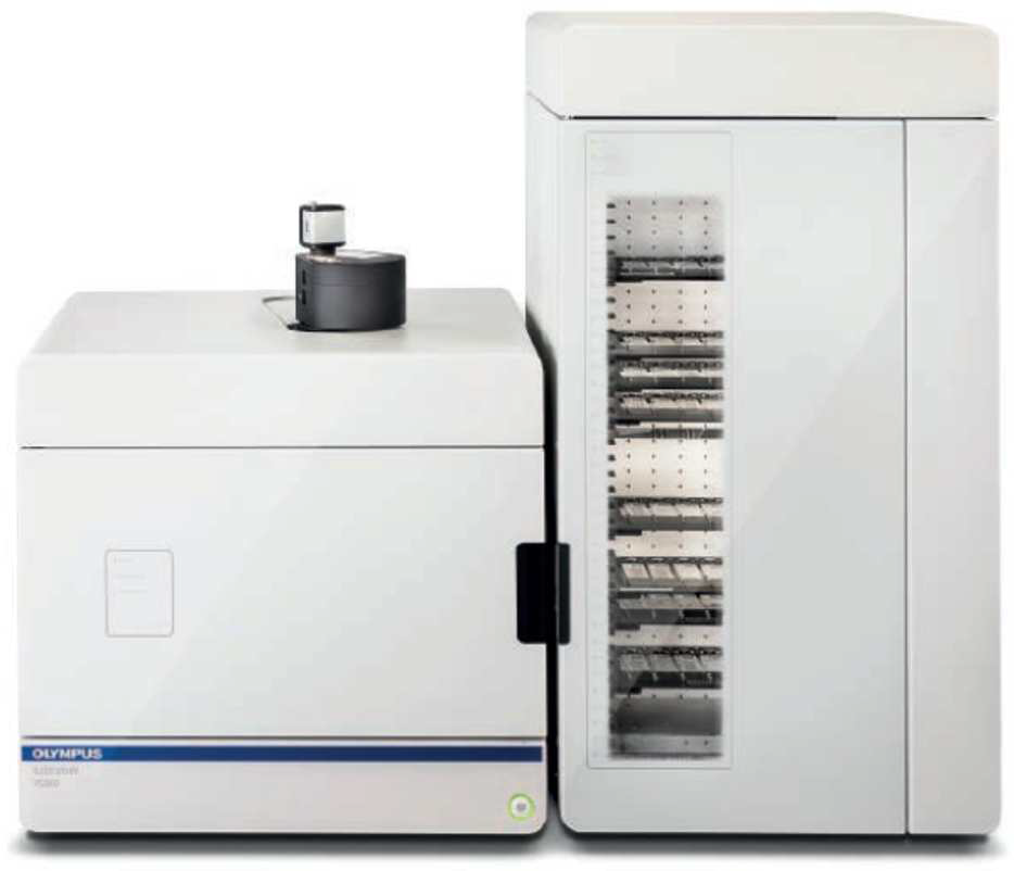
Discover Flexible Slide Scanning
The Olympus Slideview VS200 is a flexible solution for high-throughput and high-quality slide scanning. It has five different observation methods – brightfield, fluorescence, darkfield, phase contrast and polarized light – and can automatically handle a mixture of slide sizes from 26 × 76 mm to 102 × 127 mm. Fluorescence multiplexing is also supported, which makes it possible to create images with up to 32 channels with reliable and precise alignment between channels.
Outstanding Image Quality, Reliable Quantification
For crisp, analysis-ready images, the VS200 captures every detail from slides, across all five observation methods. The optical path has been designed specifically to take advantage of Olympus’ new X Line objectives, which overcome the trade-off between resolution, image flatness and color accuracy – delivering improvements in all three aspects simultaneously. Using true magnifications, from 2X to 100X, it also includes automated oil scanning while the stable and accurate focus of the VS200 gives users confidence when scanning slides for quantification purposes.
A Fluid Workflow for Every User
The VS200 is designed for users of all levels. The basic operation mode enables users to acquire images in as little as three clicks, while the expert mode provides experienced users full control, allowing them to set custom settings to different imaging projects. This is particularly useful if an imaging project requires the acquisitions of different sets of slides over a long timeframe. For ease of use and throughput, the VS200 has a robotic automated slide loader that can handle up to 210 slides, also in mixed sizes. The hot-swap function allows users to change slides during scanning, in case of urgent needs. Efficient working is natural, since the VS200 makes it possible to edit scanned images in parallel with ongoing scans. An automated barcode reader provides reliable capture and recording of information about each slide. The VS200 slide scanner software and hardware can be configured and expanded onsite: going from simple brightfield up to multiplex fluorescence scanning, with deconvolution and AI functions, allows facilities to effortlessly follow the constantly evolving application needs.
KEYWORDS: high-throughput, slide scanning, slide imaging, brain research, drug discovery
SPEAKER:
Ralf Dietzel, Omicron-Laserage Laserprodukte GmbH
ABSTRACT:
Omicron introduces its latest development LaserNest®, a single-mode desktop laser for microscopy. The presentation also includes information about the multi-wavelength laser light engines LightHUB+ and LightHUB Ultra with up to seven wavelengths.
SPEAKER:
Jan Giesebrecht, Supervisor, Product Applications Specialist, Thermo Fisher Scientific
ABSTRACT:
This live webinar will focus on the visualization and image processing of large 3D image data using the new capabilities of the Amira™ Software solution by Thermo Fisher. The head of the webinar will give you the possibility to learn, through a step-bystep demo, the recently added functions of Amira™ and its new Xplore5D extension. XPlore5D offers the fast and easy exploration of large multi-channel and time-series data. We will also highlight and demonstrate Deep Learning based training and prediction modules.
High-Speed Atomic Force Microscopy and Super-Resolution Optics – Multiparametric Correlative Microscopy Solutions from Bruker Nano Surfaces
SPEAKERS:
Dr. Tanja Neumann, JPK BioAFM, Bruker Nano GmbH,
Dimitar Stamov, JPK BioAFM, Bruker Nano GmbH,
ABSTRACT:
Atomic force microscopy (AFM) currently offers premium spatial resolution of the analysed samples while simultaneously being able to correlate topography and mechanics at near native/physiological imaging conditions. The most recent AFM developments have made it further possible to resolve single molecular dynamic processes on the millisecond scale. In turn, the combination with advanced/customised optics leverages the advantages of immunolabelling techniques for truly correlative microscopy.
During this webinar we will give an overview of correlative microscopy applications featuring fast imaging and advanced super-resolution optical setups. We will also show examples of the accuracy of registering/overlay the AFM and optical images on commercially available DNA origami structures with dimensions below the diffraction limit.
We will further introduce how some of the most recent technological developments have led to unprecedented imaging rates in fluid, setting new milestones in high-speed scanning capabilities. Bruker recently launched the fastest commercially available high-speed AFM (NanoRacer®) enabling real-time visualization of dynamic biological processes taking place on the sub-20-milisecond scale. We will introduce the concept and applications of high-speed imaging for real-time visualization of protein binding kinetics, as well as molecular and cellular dynamics.
Applying Single Molecule Localization Microscopy to Structures Deep Inside the Sample
SPEAKER:
Dr. Clemens Schneider, Sales product Specialist FM EEMEA, Bruker
ABSTRACT:
Single Molecule Localization Microscopy (SMLM) is a very widely used approach for superresolution microscopy. Compared to intensity-based superresolution microscopy images (like in STED or SIM), the localization data obtained in SMLM experiments open new ways for data processing and analysis. Having the precise 3D localization information of individual dye molecules in the sample can be used efficiently for analysis of resolution, spatial distribution, clusters, true colocalization (of clusters) and much more.
In most commercially available systems, all of these advantages are only exploited in regions close to the cover slip, since TIRF (or “dirty-TIRF”) illumination is used for reducing the background coming from out-of-focus planes. The Bruker Vutara VXL system is different in that respect. The dedicated widefield illumination in combination with the biplane detection module opens up the possibility of acquiring SMLM data more than 30 μm inside the sample.
This workshop will explain the technical requirements for performing SMLM deep inside the sample and demonstrates post-acquisition steps for SMLM data processing and analysis.
The Sample Zoo and adequate Light-Sheet Microscope Solutions from Luxendo, Bruker FM
SPEAKER:
Dr. Malte Wachsmuth, Luxendo GmbH, Bruker FM
ABSTRACT:
Light-sheet microscopy (LSFM) is today a well-known and valuable technology to gain new and deep insights of a wide variety of scientific research areas. Sample imaging across scales – with low photo toxicity and low photo bleaching effects at very high speed.
Which LSFM geometry is the most appropriate for your actual sample size, the structure you want to image, and your desired resolution? What are prerequisites to take into account to acquire the most fascinating, high content images?
Which mounting procedures are the most useful for what type of sample? Live samples, fixed samples or cleared samples, they all have specific needs and treats, and these will be addressed.
In this webinar we present an overview of our product portfolio and the key facts regarding lenses, geometries, sample size, mounting techniques, and resolution that characterizes the different specialisations of our light-sheet microscopes.
SPEAKERS:
Raino Ceccarelli, PhD., Head Of Product Development, CrestOptics
Luca Clario, Business Developer Specialist, CrestOptics
ABSTRACT:
The number of biology-driven publications that use Super-Resolution Structured Illumination Microscopy (SR-SIM) has increased significantly in recent years, indicating the usefulness of the technique as a tool for discovery. The increased use of SR-SIM also highlights the limitations of the current approaches. Revealing more biological complexity requires fast simultaneous acquisition of as many labels as possible, deep inside complex specimens. A major handicap of all SR methods is their susceptibility to aberrations, especially when imaging deeper than 10μm, which impacts contrast and resolution. To enable all researchers to overcome this multidimensional challenge, CrestOptics has developed DeepSIM, the first SR-SIM module that is compatible with all standard up-right or inverted microscopes. Despite being a high-end super resolution system (100nm in lateral X,Y and 300nm axial Z resolution), DeepSIM is as easy to operate as a confocal system – it doesn’t require any special probes and researchers can maintain their standard sample preparation protocols which guarantees a seamless evolution from Confocal to SR. We have also equipped the system with robust system calibration, pre-optimized acquisition modalities and automated data processing to make the system easier to use. We are aware that establishing SR-SIM as a ubiquitous tool for routine life science research applications requires much more than just ergonomic design and intuitive handling. With this in mind, we have developed the DeepSIM to be a truly enabling technology. Performance is combined with the flexibility of a modular, expandable system available either as standalone equipment or SR add-on module for CrestOptics X-light V3 Spinning Disk system. To further expand SR-SIM application for the study of structural dynamics and molecular interactions, CrestOptics has dedicated itself to improving live-cell imaging capabilities, despite non-trivial challenges like increasing temporal resolution and adapting to 3D imaging deep inside complex specimens. The webinar will showcase the new DeepSIM SR module and demonstrate how it can help researchers to obtain truly meaningful and contextualized deep data, even from live samples over 50μm thick.
SPEAKERS:
Marit Smeets, Delmic B.V.
Katherine Lau, Delmic B.V.
Guido Ridolfi, Delmic B.V.
ABSTRACT:
Unlocking the power of cryo-ET
Cryo-electron tomography (cryo-ET) is an extremely powerful technique that allows studies of the cellular content at the nano-scale in a near-native state. However, the current cryo-ET workflow is error-prone resulting in damaged, contaminated and devitrified samples that do not result in useful tomography data. In the workflow, there are two main challenges: keeping the sample ice contamination-free and capturing the region of interest (ROI) in the final sample.
To overcome these problems, we developed two workflow solutions called METEOR and CERES to streamline the entire cryo-ET preparation workflow. METEOR is an integrated fluorescent light microscope (FLM) that greatly enhances the ROI targeting inside the FIB/SEM while reducing the number of handling steps required. The CERES Ice Defence System consists of multiple innovative tools that are tailored to minimize ice contamination at different steps of the workflow. In this webinar we will show how using both CERES and METEOR will increase the efficiency of the entire workflow and dramatically increase the amount of high-quality cryo-ET data that can be turned into new biological insights.
Bridging the resolution gap with large-scale EM
Correlative Light Electron Microscopy is a symbiosis of techniques: fluorescence microscopy has a broad range of capabilities such as detecting specific proteins or molecules and monitoring dynamic processes in live material. EM provides data on the structural basis of these processes, by showing all membranes and structures in the sample. Combined, these two are an extremely useful research tool.
The EM part of the workflow is a bottleneck that typically forces investigators to only focus on a much smaller region of interest, compared to what can be observed with Fluorescence Microscopes. This means that unless a substantial amount of time is spent on EM, the technique is primarily qualitative.
Here we demonstrate how FAST-EM, a 64-beam electron microscope designed to drastically increase imaging throughput while maintaining high image quality, is used to collect images at nm resolution from large tissue samples. The improved throughput and automation of FAST-EM enables a shift towards using EM as a fast quantitative analysis tool. Higher throughput drastically shortens the time needed to acquire project data, and opens the door for projects of unprecedented scale, so to closing the resolution gap between EM and FM.
With FAST-EM we can easily capture with EM mode a FOV that is as large as the FM FoV (100 times faster) and collect detailed information on every labeled spot.
SPEAKER:
Bruno Sanguinetti, CTO at Dotphoton
ABSTRACT:
The combination of fast imaging techniques and machine learning are leading to scientific breakthroughs as well as high-impact applications, such as personalized medicine or autonomous driving. They are also generating vast quantities of image data, which is difficult to move around as well as expensive in time, energy and money. Image compression can help, but the usual techniques are not suitable for quantitative data.
Here we present Jetraw, a raw image compressor that preserves the metrological accuracy of raw data, and achieves compression of 8:1 at the speed of 10Gbps networks. This performance is achived through a metrological traceability to the specific image sensor and settings, which gives Jetraw images additional powers, such as physically-accurate data-augmentation and normalization, features that allow to increase, and to test, the reliability of AI algorithms.
SPEAKERS:
Henning Ortkrass*, Marcel Müller*, Anders Kokkvoll Engdahl*, Thomas Huser*,
Gerhard Holst**
*Biomolecular Photonics, Department of Physics – D3, University of Bielefeld, Germany;
**Research Department, PCO AG, Kelheim, Germany
ABSTRACT:
There is a trend in microscopes towards larger field-if-view and larger image circles to be able to receive more information with the recording of one image. For that reason camera manufacturers followed in designing and creating new scientific cameras with larger image sensors and diagonals as well. But, not only the diagonal but even more the pixel size has to be selected properly according to the applied microscope magnification. The relationship will be discussed within this webinar.
Further, it is known that all microscope systems have an optical transfer function or modulation transfer function, and so do cameras. But what is the impact on modern microscope methods like single molecule localization microscopy and structured illumination microscopy? To answer this question, a series of experiments has been done with 4 scientific cameras, which had image sensors with the same pixel size but different MTF. First results are presented within this webinar.
SPEAKER:
Dr. Kirstin Elgass, Business Sector Life Sciences, ZEISS Research Microscopy Solutions
ABSTRACT:
Since the routine use of fluorescent proteins began in the 1990s, the drive to image live samples has revolutionised our understanding of modern biology. For all live experiments, the most vital consideration is keeping light exposure to an absolute minimum since damage caused by excessive light dose can compromise the integrity of the resulting datasets.
Very often minimising light dosage means reducing the number of acquired images to decrease the length of the timelapse or the frequency of image capture. However, either of these approaches risks missing key events of interest.
Lattice light sheet technology enables 3D acquisition at subcellular resolution and with unprecedented gentleness. First published in 2014, this approach has proven itself to be extremely powerful for long-term studies and homebuilt instruments have generated numerous high impact publications. However, the scope and usability of this approach has been limited since this technology has only been available in an upright configuration and has required significant time and expertise to make the necessary alignments and optimisations for generation of the exciting results.
This has all changed with the release of the ZEISS Lattice Lightsheet 7. This platform provides lattice light sheet imaging but in a convenient, inverted configuration that is compatible with the same samples and sample preparation you would use when imaging with your confocal or spinning disc microscope.
The automatic alignments and easy workflows provided by the inverted Lattice Lightsheet 7 mean that even inexperienced users can now access this cutting-edge approach and capture 3D data of their classically mounted samples over hours and days at a time.
The accessibility of such powerful technology to non-expert users sets this approach to become the new standard in 3D long term timelapse imaging of dish mounted specimens.
Date: 5 - 8 Oct 2021
Location: Virtual
Deadline(s):
Abstract submission: Closed
Registration: Closed
Organisers:
Contact: Iva Gavran
Download event poster