Best poster prizes at ‘Organoids: modelling organ development and disease in 3D culture’
The EMBO | EMBL Symposium ‘Organoids: modelling organ development and disease in 3D culture’ took place last month, bringing together experts to discuss the ability to grow human tissues from stem cells in 3D culture that has the potential to revolutionise the drug discovery process and regenerative medicine.
The scientists presented their latest research and reveal parallels between various tissue models. Over the course of four days, 466 on-site participants and 280 virtual attendees watched 47 talks, 2 keynote sessions, and 17 flash talks across 5 sessions.
We held two poster sessions up on the helices of the Advanced Training Centre during which the presenters could discuss their research – their work was then voted for by other attendees and speakers. A whooping six best poster prizes have been awarded during the meeting and we are pleased to share them with you.
iPSC‑derived human organoid‑based model of intestinal filovirus infection
Presenter: Elizabeth Yvonne Flores, Boston University School of Medicine, USA
Collaborators: Adam J. Hume, Elizabeth Yvonne Flores, Judith Olejnik, Pushpinder Bawa, Elke Mühlberger, Gustavo Mostoslavsky

Affected Ebola virus disease (EVD) patients lose copious amounts of fluids in a matter of days, rapidly deteriorating into hypovolemic shock and death. Similar intestinal manifestations were also reported for Marburg virus (MARV) disease, another filovirus. At present, available animal models insufficiently recapitulate the gastrointestinal symptoms of EVD patients. To fill this gap, we have established an induced pluripotent stem cell (iPSC)‑derived human intestinal organoid (HIO) model that can be primed towards proximal (small intestine) or distal (colonic) intestinal lineages. Three‑dimensional tissue‑specific organoids recapitulate the heterogeneity, architecture, and cellular functions of the primary tissue, thus representing a powerful tool to study development and disease. The generation of a hiPSC CDX2‑GFP reporter line highlights the role of CDX2 as a marker for the emergence of hindgut intestinal progenitors during our differentiation protocol. This platform can facilitate the study of late‑stage EVD gastrointestinal symptoms, including diarrhea. Single‑cell RNA sequencing characterization of the HIOs revealed a significant number of CDX2 and Villin 1 expressing cells as well as separate clusters indicating the major transcriptional changes in cell identity that are known to occur during the stages of intestinal differentiation. The HIOs exhibit a distribution of the different cell types that physiologically resemble the human intestinal epithelium. We employed the generation of these organoids to study the effects of filovirus infection on intestinal epithelial integrity. Successful robust EBOV and MARV infections of hiPSC‑derived HIOs, affecting mostly epithelial CDX2+ enterocytes was achieved. The infected cells showed signs of cell damage. Transcriptomics analysis indicated the modulation of cell junction pathways and a set of ion transporters known to play a role in the induction of diarrhea. Taken together, these data suggest that EBOV and MARV compromise barrier integrity of the intestinal epithelium and cause abnormal ion flux as the basis for gastrointestinal dysfunction and diarrhea.
Spiked up microglia-containing neural organoid model
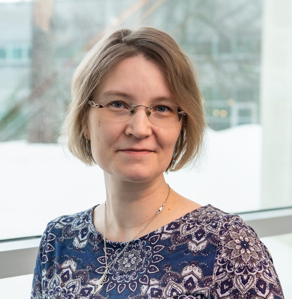
Presenter: Paula Korhonen, University of Eastern Finland
Collaborators: Mireia Gómez-Budia, Minna-Mari Tervo, Nataliia Novosolova, Nelli-Noora Välimäki, Sohvi Ohtonen, Anssi Pelkonen, Adriana Della Pietra, Anastasia Shakirzyanova, Vera Lezhneva, Susanne Michels, Raisa Giniatullina, Polina Abushik, Antonios Dougalis, Tarja Malm
Brain development is studied mainly in animals due to unavailability of human brain cells. However, human and mouse brain differ from each other significantly. Human induced pluripotent stem cells (iPSC) offer a new way to get an unlimited access to human brain cells, and iPSC-derived cerebral organoids a promising platform to study neurodevelopment and associated pathologies. We have shown in our novel microglia-containing organoids (iORGs) that microglia boost neuronal activity during the brain development. In this study, we have optimized our model to enhance neuronal maturation further and carried out in depth electrophysiological characterization over the 7-month culture period. iORGs were grown from 5 iPSC lines and neuronal differentiation was induced using a pattern of growth factors. iORGs were sliced with a vibratome into 500 µm thick slices, placed on inserts and grown as air-liquid interphase (ALI) culture. Microglial progenitors were incorporated on top of the slice. Immunohistochemical stainings were carried out at different timepoints (4, 5, 6 and 7 months) to confirm the presence of neuronal cell types and microglia. Multi-eletrode array (MEA) measurements were carried out using 3D electrodes in MEA2100-Mini-60-System. To map neuronal excitatory characteristics, N-methyl-D-aspartate (NMDA) was applied and spikes and local field potentials were measured. Patch clamp technique in current and voltage clamp modes was used to record basic neuronal parameters, action potentials and spontaneous excitatory and inhibitory currents. Our data revealed a high variability in the maturation potential between hiPSC lines. Microglia accelerated neuronal maturation and decreased NMDA-induced neuronal network hyperexcitability. We also confirmed inhibitory GABA response at later timepoints (7 months) indicating that developmental GABA switch from excitatory to inhibitory occurs in our model, supported by increase in parvalbumin-positive interneurons over time. To conclude, we have an electrophysiologically relevant model recapitulating neuronal maturation in a time-dependent manner. Our iORGs can be used e.g. in the investigation of genetic factors or environmental exposures and they provide a way to study cell-cell interaction in human context.
View poster (some parts of the poster have been blurred due to unpublished data)
Effect of Fibroblasts on the Structure of Human Hair Follicle Stem Cells-Derived Hair Follicle Organoids
Presenter: Weiling Lian, Fudan University Huashan Hospital, China

Develop advanced preclinical models for androgenetic alopecia (AGA) by incorporating hair follicle fibroblasts (HFFBs) into hair follicle stem cells (HFSCs) derived hair follicle organoids. Investigate the role of HFFBs in organoid differentiation and maturation through epithelial-mesenchymal interactions (EMI). Primary HFSCs and HFFBs from AGA patients’ hair follicles were isolated and characterized. HFSCs were cultured as 3D spheroids, differentiating into HFSC-derived organoids (HFSCOs). HFFBs were added to create HFSCs and HFFBs-derived organoids (HFSCFBOs). Organoids were analyzed with microscopy, H&E staining, and immunofluorescence. Cytokine microarray sequencing and related signal inhibition explained HFFBs’ roles and pathways. HFSCs showed robust proliferation and stemness gene expression. HFFBs exhibited stable proliferation and increased dermal papillae gene expression. During the 1-week incubation, HFSCOs and HFSCFBOs exhibited hair follicle-like structures, with 1 hair follicle unit inducing an average of 137.8 – 708.8 organoids. The participation of HFFBs can improve the proliferation. Immunofluorescence confirmed marker expression including KRT15, CD200, LHX2, P-cadherin, SOX2, VIMENTIN, KRT6, KRT81, ASMA, TRP1, and PMEL in HFSCFBOs on 7th day and even after 60 days. And the expression has structural distribution characteristics, compared to HFSCOs. Cytokine analysis revealed that HFFBs may promote epidermal growth factors production and up-regulated IGF-1R, Wnt/β-catenin, and MAPK/p38 signaling pathways. High expression of IGF-1R, Wnt and MAPK signals were detected in HFSCFBOs. And inhibiting IGF-1R, Wnt and MAPK blocks HFSCFBO differentiation. HFSCs differentiated into hair follicle organoids, with HFFBs enhancing structure and functionality. The study highlights EMI and the niche microenvironment’s role in organoid differentiation. This hair follicle organoid provides a theoretical basis for precision medicine and hair regenerative research.
Due to the confidentiality of the unpublished data, we cannot share the poster.
Human neural organoids beading it: neurons in 3D culture display signs of injury following mpox virus infection
Presenter: Isabel Schultz-Pernice, University of Bern, Switzerland
Collaborators: Amal Fahmi, Yen-Chi Chiu, Blandina I. Oliveira Esteves Criblez, Teodora David, Antoinette Golomingi, Beatrice Zumkerh, Damian Jandrasits, Roland Züst, Selina Steiner, Carlos Wotzkow, Fabian Blank, Olivier Engler, David Baud, Marco P. Alves

Early in May 2022, first cases of mpox virus (MPXV) infection without traceable contact to African population or fauna were reported in UK. Since then, 113 countries registered cases of infection, marking the largest outbreak outside of endemic regions. First manifestations of mpox include fever, lymphadenopathy, and muscle aches, followed by vesiculopapular rash development. Neurological manifestations, ranging from mild headache to fatal encephalitis, may develop in around 3% of patients. In September 2022, MPXV DNA was detected in the cerebrospinal fluid of a young, previously healthy woman affected by encephalitis, suggesting neuroinvasive potential of MPXV. Despite reports of deadly encephalitis cases date back to 1987, mechanisms driving acute neurological manifestations during MPXV infections have been poorly investigated. Using human neural organoids (hNOs) we explored the susceptibility of cerebral tissue to infection with a patient‑derived 2022 isolate of MPXV. MPXV efficiently replicates in hNOs as indicated by the exponential increase of viral loads upon infection and the elevated frequency of MPXV‑positive cells over time. Transmission electron microscopy imaging of infected hNOs confirmed the presence of mature viral particles as well as perinuclear viral factories. We additionally confirmed susceptibility of different cell types to the virus, including glial cells and neurons. Furthermore, we detected the presence of viral antigen in neurites and in foci of grouped cells distributed throughout the tissue. In line with this, comparing released and cell‑associated MPXV titers, we observed significantly more cell‑associated infectious virus, spanning over several orders of magnitude, suggesting spread of MPXV by cell‑to‑cell contact. While hNOs displayed no evident morphological changes, we observed spheroid formation on neurites, indicative of neuronal damage. Cell injury was further confirmed by the detection of apoptotic marker cleaved caspase‑3 within neurite spheroids. Our findings underline the value of hNOs to model neurotropic virus infection in the human host and underline the need to shed light on the mechanisms driving severe complications following infection by this previously neglected pathogen.
Human intestinal organoid co-culture model with tissue-derived immune cells uncovers novel immune-epithelial interactions
Presenter: Michelle Steinhauer, BioMed X Institute, Germany
Collaborators: Inga Hensel, Szabolcs Éliás, Martin Resnik-Docampo

Intestinal epithelial cells function as a protective barrier towards potential harmful luminal content. They release immune regulators to attract basally residing immune cells to initiate immune responses and control inflammation. Loss of intestinal barrier integrity, and alterations in intestinal immune cell function is correlated with gut dysbiosis, cancer and inflammation processes like e.g. autoimmune diseases. However, interactions between lamina propria immune cells and intestinal epithelial cells are poorly understood due to the lack of a relevant in vitro model. We established and characterized a near‑physiologic, reproducible 3D co‑culture model that for the first time utilizes human lamina propria‑derived immune cells in co‑culture with donor‑matched intestinal organoids to specifically study immune‑epithelial cell interactions. A time‑dependent increase in interacting immune cells was observed using live‑cell imaging, accompanied with an increase in IL‑6, IL‑18, IL‑8, and MCP‑1 after 48h in co‑culture supernatant. Using spatial phenotyping we could identify dendritic cells as well as B and T cells, interacting with organoids. Single cell RNA sequencing of human colon co‑culture revealed transcriptomic changes in distinct epithelial and immune cell clusters. In summary, our work provides the first near‑physiologic primary human co‑culture model that not only enables us to better understand the intestinal immune‑epithelial cell biology but also serves as a platform to study diverse pathologic conditions with the potential to be used for drug discovery processes.
Collective organization of circadian rhythms in murine intestinal organoids
Presenter: Elena Tonin, EPFL Switzerland
Collaborator: Felix Naef
The circadian clock orchestrates physiological and behavioral rhythms in living organisms with a period of approximately 24 hours. This vital adaptation on Earth allows mammals to compartmentalize biological processes at different times of the day. Disruption of these rhythms has deleterious consequences, for example on tissue regeneration and cancer progression. However, there is a lack of understanding of how the molecular clock, cell differentiation and cell cycle integrate during homeostasis and disease. Also, circadian research is mainly conducted in vivo or in homogeneous populations of cells with some results suggesting that the clock may ‘gate’ the cell cycle to certain time windows, while others have shown that cell cycle progression influences the clock. To shed light on the topic, we implemented the fast‑proliferating multicellular system of mouse intestinal organoids to study the collective behavior of circadian rhythms during proliferation and differentiation, with quantitative and mathematical analysis. Few studies reported circadian oscillations in intestinal organoids and addressed the interaction with the cell‑cycle, although without considering the circadian properties of different cell‑types in the mammalian epithelium.
For this reason, with the help of microfabricated hydrogels, CRISPR‑editing as well as bioluminescent and fluorescent imaging, we are disentangling clock dynamics at the single cell level during homeostasis and regeneration. We are able for the first time to show single cell resolution of circadian rhythms in a multicellular tissue. In particular, microarrays of epithelial tissue show overall robust synchronized circadian oscillations but highlight variable cell type‑specific clock amplitudes. Additionally, it seems that also the fast‑proliferative stem cells possess circadian oscillations. This project contributes to setting the basis for in vitro multicellular circadian studies that will help to understand the current difficulties in treatment of neoplasms and regeneration.
Due to the confidentiality of the unpublished data, we cannot share the poster.
Find out more about the #EESOrgan meeting from the blog post written by Elisa Heinzelmann, who participated as an event reporter: https://www.embl.org/about/info/course-and-conference-office/2023/11/my-journey-through-the-embo-embl-symposium-organoids-modelling-organ-development-and-disease-in-3d-culture/
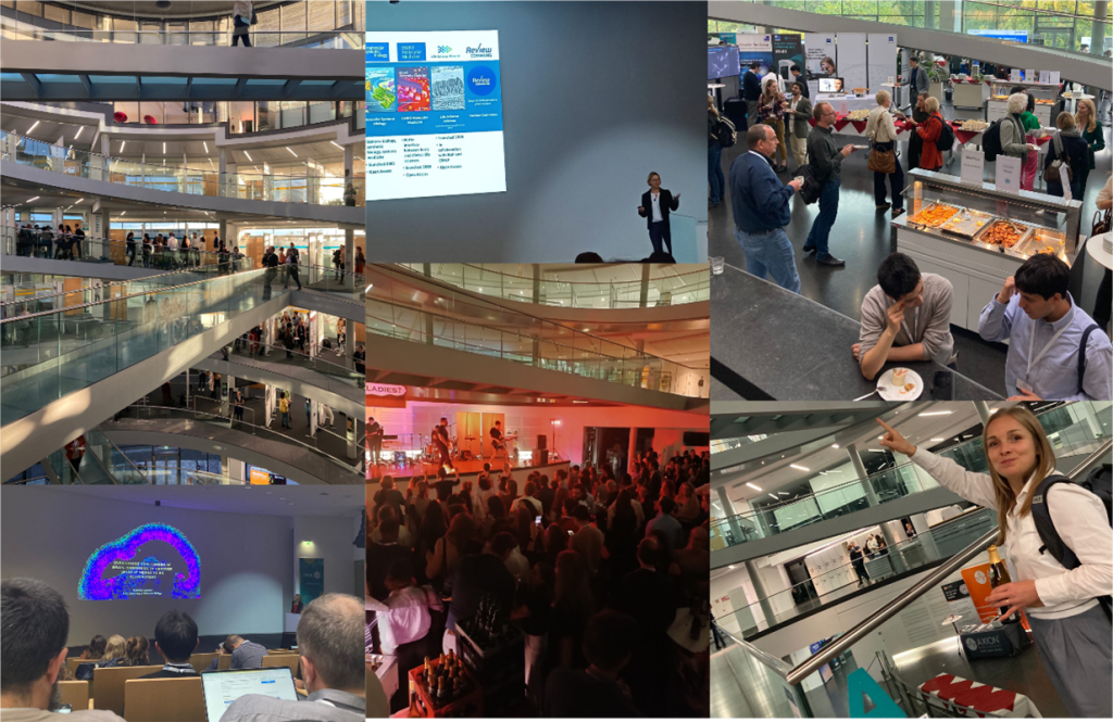
The EMBO | EMBL Symposium ‘Organoids: modelling organ development and disease in 3D culture’ took place from 18 – 21 October 2023 at EMBL Heidelberg and virtually.