Poster prizes at ‘Defining and defeating metastasis’ – meet the winners!
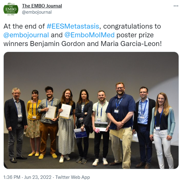
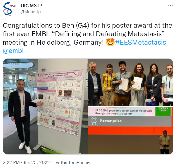
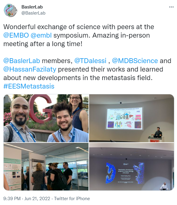
We are excited to present the poster prizes awarded at the recent EMBO | EMBL Symposium ‘Defining and defeating metastasis’, hosted at the Advanced Training Centre in Heidelberg and bringing together researchers from diverse fields to enhance our understanding of the dissemination and metastatic colonisation of tumour cells. It provided a unique opportunity for interdisciplinary exchange on current approaches and future collaborations on metastasis and its therapeutic challenges. As with most events this year, for many participants this was the first onsite meeting that they attended since early 2020 which made it very special. It was a fantastic opportunity to meet in person for the three days full of exciting science, exchanging ideas, presenting latest research, catching up with old friends and making new ones. There were two live poster sessions during which the presenters could discuss their research– their work was then voted for by other attendees and speakers. We are pleased to be able to share with you the research from four out of six winners of the conference prizes: congratulations to all!
Jagged-1 promotes breast cancer metastasis through the lymphatic system
Presenter: Benjamin Gordon

Abstract
While early detection of breast cancer (BC) has improved prognoses, there is an urgent need to improve outcomes for patients with distant metastatic disease. Higher expression of the Notch ligand JAG1 in primary BC tumors is strongly associated with lymph node metastasis and patient mortality, but causality is unclear. We show that JAG1 expression is higher in metastatic BC cells colonizing lymph nodes than in primary tumors, suggesting that tumor cells with high JAG1 are preferentially able to metastasize to lymph nodes. JAG1 expression is higher in a derivative of BC line MDA MB 231 selected for tropism to lymph nodes (MDA231 LN) than in the parental line or derivatives with other tropisms. To determine the mechanism(s) of JAG1 mediated metastasis, we generated clonal JAG1 knockout cell lines from MDA231 LN cells with corresponding JAG1 rescue lines. We investigated the role of JAG1 in spontaneous metastasis under clinically relevant conditions by orthotopically implanting JAG1 knockout and expressing cells, resecting the primary tumor, and following long term metastatic spread in a mouse model. Quantification of tumor cells in blood showed that survival, metastatic burden, and JAG1 expression did not correlate with the number of circulating tumor cells. Conversely, JAG1 expression drove an increase in lymph node and body wide metastatic burden and a trend toward decreased survival. In this model, metastatic cells were abundant throughout lymph vessels, suggesting lymphatics are the primarily route of dissemination. Preliminary transcriptional analysis suggests that JAG1 alters interactions with lymphatic endothelial cells (LEC), leading us to examine downstream events in co cultures of LEC with lymphatically invasive BC lines. Deciphering tumor lymphatic endothelial signaling events may open new avenues to target BC metastasis.
Poster Prize from the EMBO Journal
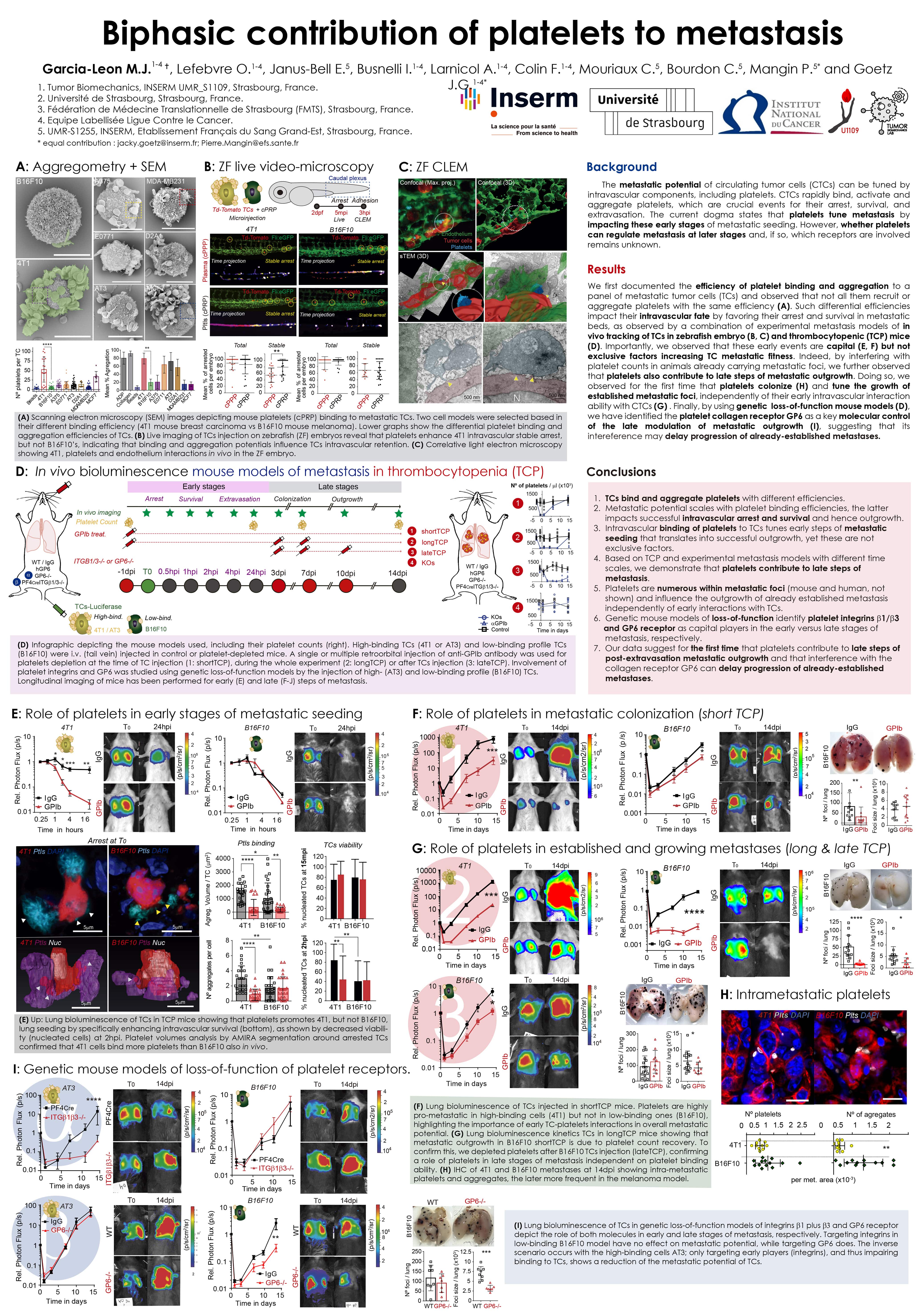
Biphasic contribution of platelets to metastasis
Presenter: María J. García-León

Abstract
Metastasis still remains elusive to treatment, with an overwhelming mortality rate of 90%. Accumulating evidence indicates that metastatic potential of circulating tumor cells (CTCs) can be tuned by intravascular components, including platelets. Platelet depletion impairs metastasis, which can be rescued upon platelet transfusion. Mechanistically, CTCs rapidly bind, activate and aggregate platelets, events that are crucial for the arrest, survival, and extravasation of the former. The current dogma states that platelets tune metastasis by impacting CTCs behaviour at early stages of metastatic seeding. However, whether platelets can regulate metastasis at later stage and which receptors may be involved remains unknown. In this study, we first documented the efficiency of platelet binding to a large panel of metastatic TCs and observed that not all recruit or aggregate platelets with the same efficiency. Interestingly, such binding impacts their intravascular fate by favoring their arrest, as observed in a combination of experimental metastasis models in thrombocytopenic (TCP) mice and zebrafish embryo. Using longitudinal imaging of metastatic seeding and growth in TCP mice at unprecedented spatial and temporal resolution, we demonstrated that binding and aggregation correlates with their metastatic potential in vivo. Additionally, by the dynamic in vivo tracking of TCs in the lungs of fully TCP mice, and the quantification of platelets depositions around arrested CTCs at seeding and late metastatic outgrowth, we showed that early platelet binding, aggregation, clot formation, and the subsequent increased adhesion and survival at lung microvessels, are capital but not exclusive factors increasing TC metastatic fitness. We observed that platelets contribute to late steps of metastatic outgrowth by experimentally interfering with platelet counts in animals already carrying metastatic foci. Doing so, we observed that platelets tune the growth of established foci, independently of their early intravascular interaction with CTCs. Finally, we have identified the platelet collagen receptor GPVI as key in this late modulation of metastatic outgrowth, suggesting its targeting in specific cancer types as a promising adjuvant therapy in oncologic patients to stop the metastatic progression.
Poster Prize from EMBO Molecular Medicine
Colonic fibroblasts in tissue homeostasis and cancer
Presenter: Michael Brügger

Michael Brügger – University of Zürich, Switzerland
Abstract
Colorectal cancer (CRC) is among the most prevalent cancers in Switzerland (2nd in women 3rd in men, BFS statistics 2013 2017) and worldwide (3rd in women and men). More than half of the patients diagnosed with CRC either harbour metastases or will develop metastatic disease, which is the primary cause of death for CRC patients. There is therefore a dire need for new therapies. These must be guided by a better understanding of the metastatic process. We are only now starting to appreciate the contribution of not only tumour cells themselves, but also the non tumour stromal cells of the tumour microenvironment (TME) to tumour growth, progression and metastasis. To understand how non tumour stromal cells are changed in CRC it is integral to first characterize their identity and functions during colonic homeostasis.
To describe the stromal cell populations in an unbiased manner, we carried out a single cell transcriptome analysis of the adult murine colon, producing a high quality atlas of matched colonic epithelium and mesenchyme. We identify two crypt associated colonic fibroblast populations that are demarcated by different strengths of platelet derived growth factor receptor A (Pdgfra) expression. Crypt bottom fibroblasts (CBFs), close to the intestinal stem cells, express low levels of Pdgfra and secrete canonical Wnt ligands, Wnt potentiators, and bone morphogenetic protein (Bmp) inhibitors. Crypt top fibroblasts (CTFs) exhibit high Pdgfra levels and secrete noncanonical Wnts and Bmp ligands. While the Pdgfralow cells maintain intestinal stem cell proliferation, the Pdgfrahigh cells induce differentiation of the epithelial cells. Notably, these cell populations are conserved in the human colon.
Recently, we established a murine model of metastatic colorectal cancer, based on the orthotopic endoscopy guided injection of cancer organoids (colonic organoids harbouring mutations in APC, Kras, Tp53 and Smad4). In this context we study how the abovementioned fibroblast populations are affected by the primary tumour and how they in turn affect tumour progression.
Single cell transcriptomic profiling of brain metastatic founders in small cell lung cancer patient derived models to identify potential vulnerabilities
Presenter: Maria Peiris-Pagès
Abstract
Background: Brain metastasis is a major cause of patient morbidity and mortality in small cell lung cancer (SCLC) with an ~80% incidence during disease progression, contributing to the dismal 5 year survival rate of <7%. Mechanisms underpinning SCLC brain metastasis are understudied due to scarcity of brain biopsies and preclinical models. We have developed a biobank of >60 circulating tumour cell (CTC) derived patient explant models of SCLC in immunodeficient mice (CDX) where brain metastasis is routinely observed upon resection of the subcutaneous (S.C) tumour
Methods: We developed an in vivo S.C tumour resection workflow in brain tropic CDX3P to isolate single CTCs, early brain founder tumour cells and subsequent established brain metastases. Following FACS of CDX cells from dissociated mouse brain (using a human CD147 antibody) we performed single cell RNA sequencing (scRNAseq) to reveal potential molecular regulators hypothesised to support brain metastatic founding and subsequent colonisation
Results: Brain metastases were detectable in CDX3P on average 195 days after S.C implantation (study length 174 230 days). We analysed 58 single CTCs (n=6 mice, 191 230 days) and 214 brain metastatic founder cells (n=2 mice, 205 218 days) by scRNAseq. Bioinformatics analyses defined transcriptomic features underpinning single cell heterogeneity and identified sub populations within CTCs and metastatic founders indicative of brain tropic CTC sub clones. We also characterised molecular features unique to brain founders as candidates that could serve as therapy targets
Future Tissue expression of candidate genes of brain metastatic founding will be validated in CDX and patient samples. Genetic manipulation of CDX cells ex vivo combined with pharmacological approaches will be used to explore their roles in metastatic seeding and to identify potential vulnerabilities. Transcriptomic analysis of cells from established brain metastases obtained from the above in vivo protocol will be conducted to explore molecular programs of brain colonisation. Combined, these data will contribute to our long term goal of identifying novel therapeutic strategies that may ultimately improve the quality of life for the significant number of patients with SCLC who present with or subsequently develop brain lesions.
Due to the confidentiality of the unpublished data, we cannot share the poster.
Effective treatment of colorectal peritoneal metastases by exploiting a molecular subtype specific vulnerability
Presenter: Sanne Bootsma

Sanne Bootsma, Amsterdam UMC, The Netherlands
Abstract
In colorectal cancer, peritoneal metastases (PMs) associate with severe morbidity and dismal prognosis. Given the incidence of this disease and the lack of adequate treatments currently available, PMs pose a large unmet clinical need. Although PMs can be accompanied by more widespread metastatic disease, it often occurs as the only sign of dissemination. This implies that the route of metastatic spread to the peritoneum differs from that to distant organs. PMs are thought to result from cancer cells that spill into the abdominal cavity, and are able to attach to the peritoneal lining and form tumor deposits. This cascade places specific demands on the cancer cells.
Here, we report that colorectal cancers that present with PMs almost universally classify as consensus molecular subtype 4 (CMS4). This previously recognized disease entity is characterized by mesenchymal features, poor prognosis, and resistance to therapies currently used against peritoneal metastases, which explains their limited efficacy. By leveraging disease models that capture CMS4 specific features, including the ability to form PMs in vivo, we identified elesclomol as a highly effective agent. Elesclomol kills cancer cells in a copper dependent fashion by targeting the oxidative phosphorylation machinery, which we found to be a specific vulnerability of CMS4 cancers. Elesclomol Cu2+ was effective following only minutes of exposure to CMS4 cell lines and organoids, supporting its use in intra abdominal treatment procedures. It is therefore a promising candidate for the local treatment of peritoneal metastases of colorectal cancer.
Poster Prize from Metastasis Research Society
The remaining prize was:
Short talk Prize from Metastasis Research Society: Eric Rahrmann – University of Cambridge, UK
Congratulations to all six winners!
The EMBO | EMBL Symposium ‘Defining and defeating metastasis’ took place from 19 – 22 June 2022 at EMBL Heidelberg and was streamed online for virtual participants.