Best Poster and Artwork Awards — VIZBI 2021: Visualizing Biological Data
The 11th international meeting on Visualizing Biological Data, best known as VIZBI, was held virtually this year. The conference was as exciting as always, filled with great discussions, an outstanding speaker line-up and of course amazing, beautiful visuals.
The participants had the chance to vote for their favourite scientific poster and artwork — a very tough choice as all of the works were truly amazing! Here, we present you the winners.
Best scientific poster
Building a whole cell in 3D
by Martina Maritan (The Scripps Research Institute, USA) Ludovic Autin, Jonathan Karr, Markus Covert, Arthur Olson, David Goodsell.
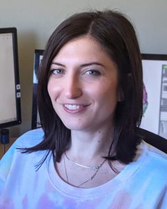
Mesoscale 3D models are powerful tools for exploring structural data across the entire range of scales, from the molecular to the cellular level. We built structural mesoscale models of a whole Mycoplasma genitalium (MG) cell with the CellPACK suite using data generated from a whole-cell MG simulation. 3D models integrate structural details into a computational model of MG, highlighting specific properties of the ingredients, and creating snapshots of the cell at defined time points of the simulations. Our modeling process goes through three steps. Firstly, we assemble a recipe: a list of all the proteins of Mycoplasma associated with a structural representation. Secondly, we create a model of the genome with DNA, RNA, RNA polymerase, mRNA, and ribosomes, with user-defined location of RNA polymerase and length of transcripts. Thirdly, we assemble the nucleoid, soluble, and membrane ingredients, and relax the whole system to resolve steric overlaps. The result is a framework for interactive construction of atomic resolution mesoscale models describing a spatial view of a whole bacterial cell. Our models are the first atomistic representation of an entire bacterial cell.
View Martina Maritan’s poster
Watch lightning talk
Second best scientific poster
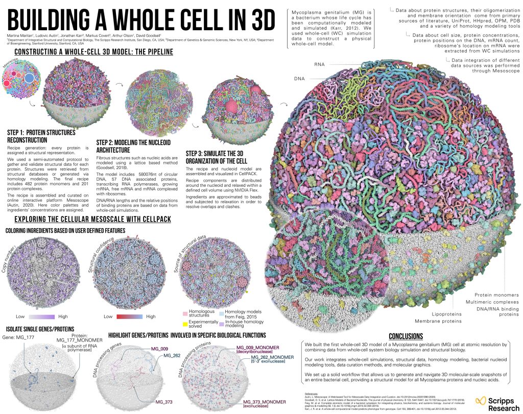
How to communicate cell behaviours visually
by Christian Stolte, Cellarity, USA.

Cellarity is pioneering a new approach to drug discovery, treating disease at the level of the cell as opposed to a single molecular target. Combining unique expertise in network biology, high-resolution single-cell sequencing data, and machine learning, the result is a new understanding of the cell’s trajectory from health to disease, and how cells relate to one another in tissues. The cell and its network of transcripts and proteins offer a more complete view of the complexity of human biology than any individual molecular target. To help communicate this, we use visualizations resembling a cityscape called ‘Cellarity maps’. Based on the UMAP dimensionality reduction technique, they use the third dimension (height) to show density. This creates landscapes where we can now use colour to encode additional dimensions, and make it easier to see different ‘cell behaviours.’
View Christian Stolte’s poster
Watch lighting talk
Best artwork
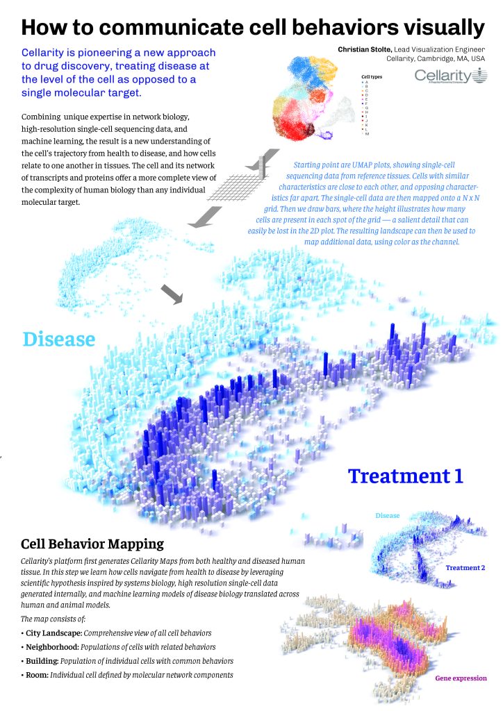
10 Hallmarks of cancer
by Karolína Kryštofová, Institute of Biophysics of the Czech Academy of Sciences, Czech Republic.

A whimsical series of illustrations inspired by the hallmarks of cancer as described by Weinberg & Hanahan in their paper Hallmarks of cancer: the next generation (2011).
View Karolína Kryštofová’s artwork
Second best artwork
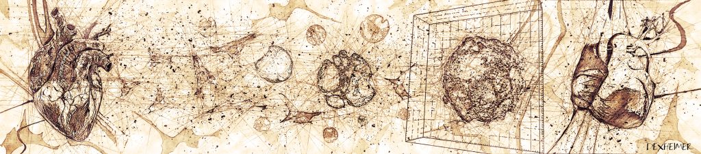
The human heart
by Philipp Dexheimer, Research Institute of Molecular Pathology, Austria.

Inspired by Leonardo Da Vinci’s original way of depicting his science and ideas, this digital painting illustrates the 21st century research process to understand formation of such a delicate organ as the human heart. Cells are derived, self-organize into 3-dimensional organoid structures, and allow unique insight into heart development and physiology. Illustrating research described in: Hofbauer et al., BioRxiv, 2020 – Cardioids reveal self-organizing principles of human cardiogenesis.”
View Philipp Dexheimer’s artwork
If you’d like to take a look at all of the posters presented at VIZBI 2021, you can! Visit the poster gallery, dive into the science, enjoy the beautiful images and be amazed by the scientists’ visualization skills.