New 3D light microscopy method reveals new perspectives on the dynamics of the cell’s skeleton
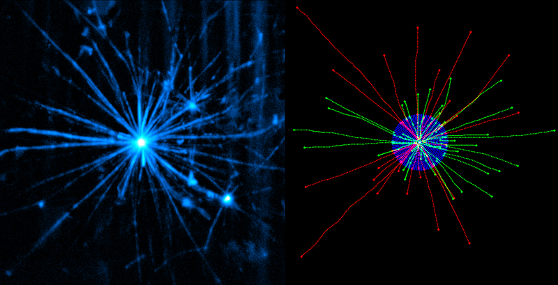
Scientists from the European Molecular Biology Laboratory (EMBL) have developed a new method to prepare and image biological samples in three dimensions with laser light-sheet based fluorescence microscopy. The technological advance, which is published in the current online issue of Nature Methods, allows for the first time the observation of the intrinsic dynamic properties of microtubules. These constitute a major part of the cell’s skeleton and can now be observed in a mechanically unconstrained and at the same time physiologically relevant context.
Microtubules form a network of protein filaments, which constantly grow and shrink. This network’s behaviour is controlled by the intrinsic properties of many different proteins. Conventional microscopy studies microtubules and other filaments in artificial set-ups, which biases certain behaviour and introduces artefacts via the hard and flat surfaces of the surrounding glass chamber. Ernst Stelzer and his colleagues Philipp Keller and Francesco Pampaloni at EMBL have managed to overcome the limitations of traditional microscopy and discovered different microtubule behaviour in an unrestricted environment, resulting in a highly accurate characterisation of intrinsic microtubule dynamics in a close to life context.
The new method is currently being adopted to study other dynamic cellular processes. The authors expect their results to have a major impact on the understanding of the mechanical properties of tissue cells. This will also influence current nanobiotechnology and have a significant effect on cancer research.


