
The eye of science
Parisa Kakanj, a postdoc in the Leptin group at EMBL Heidelberg and the University of Cologne, recently developed and published a technique to study wound healing in living fruit fly larvae. The larvae already have highly developed organs, yet their body is still transparent, which allows scientists to observe organs inside the living animal under the microscope.
When Parisa’s sister Mona – who is a visual artist – saw the images her sister created as part of her study, she realised that they could be repurposed to create a work of art.
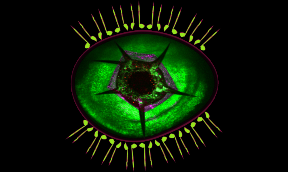
The image visible here is a composite of lateral pentascolopidial organs, a wing imaginal disc pouch, and an epithelial wound in a Drosophila larva. The organs are arranged here like eyelashes. Cells surrounding an epidermal wound appear as the iris and pupil of this artistic eye.
Credit: Parisa Kakanj, Mona Kakanj/EMBL
If you have a stunning picture of your science, your lab or your site, you can submit it here.


