Read the latest Issue

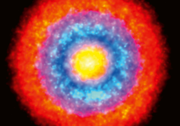
This Picture of the Week is actually more than just one image: it has been composed from thousands of individual super-resolution microscopy images. This composite image, created by Markus Mund in the Ries Group at EMBL Heidelberg, shows the radial organisation of proteins on the cell membrane during endocytosis.
Endocytosis is the cellular process in which substances are brought inside the cell. The material to be transported is surrounded by an area of cell membrane. This membrane buds into the cell to form a vesicle containing the ingested material.
This super-resolution microscopy image by Mund shows how a set of cytosolic proteins guide actin polymerisation locally. Actin patches will generate the mechanical force required to bend the cell membrane and form endocytic vesicles.
In this image, three proteins are stained: Bbc1 in red, Las17 in blue and Sla1 in yellow. Other colours represent the overlap between different rings. Bbc1 and Sla1 confine Las17 to this well defined blue ring, which triggers actin polymerisation.

If you have a stunning picture of your science, your lab or your site, you can submit it to: mathias.jaeger@embl.de.
Looking for past print editions of EMBLetc.? Browse our archive, going back 20 years.
EMBLetc. archive