Jan Ellenberg
EMBL Heidelberg
Germany
EMBL Conference
Please note that this event will now take place virtually.
The event is jointly organised by EMBL, ZEISS, The Francis Crick Institute and the VIB Ghent.
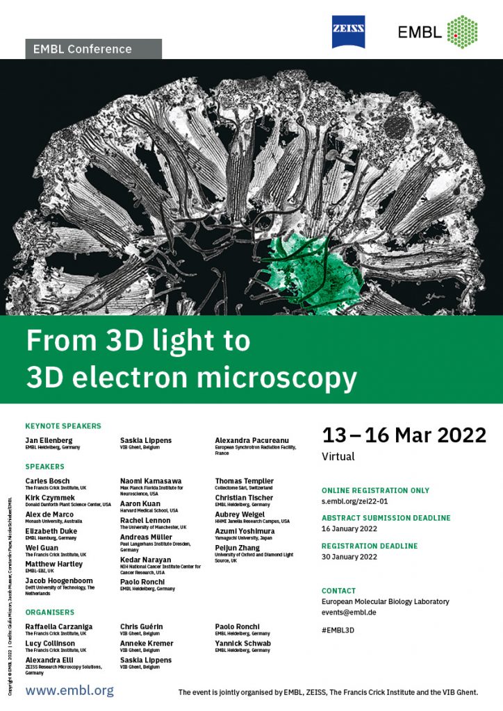
Life happens in 3D – and understanding biological context in the three dimensions has been a major driver in the past decades in both light and electron microscopy. Today the trend towards 3D continues with large volume imaging and highest resolution with modern light and electron microscopy-based techniques now enabling previously impossible insights into functional and structural biology. Correlative microscopy can combine a large variety of different imaging techniques such as confocal microscopy, X-ray microscopy and different volume EM methods e.g. Array Tomography or Serial block face imaging. Comprehensive workflow solutions and automatisation have eased the path for the use of Correlative Microscopy and volumetric imaging.
An emerging challenge resulting from the improved connectivity between the individual imaging modalities is the handling of larger and thus more complex data. Workflow flexibility, file compatibility and big data handling are the new challenges to be tackled, not only from a hardware perspective but also for image processing and visualisation.
This meeting is centred on scientific sessions covering a broad range of correlative workflows, volume EM imaging, image processing techniques as well as segmentation and visualisation. The meeting will take place from 13 to 16 March 2022 – please save this date to your calendar! Don’t miss this exciting opportunity for exchange with researchers and industry to explore the new and emerging technologies and methods for correlative microscopy and volume EM in life sciences.
Due to the unpredictable COVID-19 situation, we are going to offer the 6th Joint Workshop and Symposium ‘From 3D Light to 3D Electron Microscopy’ as a virtual event.
All participants are able to present their own scientific work in a poster presentation or an oral presentation. The organising committee will review abstracts for the oral presentations.
We look forward to meeting you virtually!
During this workshop, we will share with participants experiences about sample preparation for the different techniques presented during the conference. We will show tips and tricks for sample preparation and mounting on different supports for 3DEM and CLEM. We will cover the techniques for SBEM sample prep, FIBSEM sample prep (with and without fluorescence preservation) as well as array tomography.
Contributing people: Christel Genoud, Jean Daraspe, Olivia Muriel Lopez (all University of Lausanne)
See how non-destructive ‘Array Tomography’ (AT) works: This workshop will cover different serial sectioning approaches, strategies for image acquisition, through to aligning the 2D images to be able to generate a 3D model. These volume EM datasets can be correlated with 3D light microscopy datasets of the same sample, acquired previously by either confocal imaging of the sample live or fixed, or widefield imaging of the serial sections.
Contributing people: Jemima Burden (University College London), Ian White (University College London), Raffaela Carzaniga (The Francis Crick Institute), Stephen Furzeland (Carl ZEISS)
We will show you how XRay images can be used to determine the position of pollen tetrads in the arabidopsis anther and how to use this data to trim the sample block to be able to relocate this position in SBF-SEM for imaging of specific pollen tetrads.
Contributing people: Anneke Kremer (VIB Ghent), Saskia Lippens (VIB Ghent), Marianne Beckwith (EMBL Heidelberg), Kimberly Meechan (EMBL Heidelberg)
Learn how you can combine cryo-confocal imaging, cryo-lamella preparation and cryo-electron tomography to get high-resolution information of your favorite structure. The correlative workflow connects a confocal fluorescence microscope, with a focused ion beam – scanning electron microscope for targeted lamella preparation and a high-end transmission electron microscope. You will learn about the hardware and software involved to investigate your samples in their native state.
Contributing people: Anna Steyer (EMBL Heidelberg), Simone Mattei (EMBL Heidelberg), Zhengyi Yang (EMBL Heidelberg), Andreas Schertel (Carl Zeiss Microscopy GmbH)
This workshop will present a correlative 3D light and electron microscopy workflow that integrates in-resin confocal fluorescence microscopy, 2-photon branding and focused ion beam scanning electron microscopy (FIB-SEM), as described in Ronchi et al., 2021. We will discuss how to preserve fluorescence during sample preparation for volume EM and show how to target the FIB-SEM acquisition of specific cells of interest based on a fluorescence 3D map acquired by confocal imaging of the block.
Contributing people: Paolo Ronchi (EMBL Heidelberg), Giulia Mizzon (Heidelberg University), Manuel Gunkel (EMBL Heidelberg), Aliaksandr Halavatyi (EMBL Heidelberg), Karel Mocaer (EMBL Heidelberg)
We will present the typical serial section acquisition workflow with a multibeam scanning electron microscope, where navigation and ROI definition is routinely done on a correlated wide-field light microscope overview image of the sample. A semi-automated Experiment Wizard guides the user step-by-step through the workflow setup, emphasizing the ease-of-use required for a true high-throughput imaging experience. Besides that, operation principle and image data structure of the ZEISS MultiSEM will be introduced.
Contributing people: Anna Lena Eberle, Tomasz Garbowski, Stefan Nickell (all Carl Zeiss MultiSEM GmbH)
Hosted by Lucy Collinson and Yannick Schwab
The discussion will be moderated by international panellists all involved in community efforts around vCLEM. They will share their views and trigger feedback from the audience around 3 topics: how international communities organise access to infrastructures (open facilities, funding mechanisms); how they develop their training and outreach activities; how they help scientists with dealing with their data (standards, repositories).
Panel members:
Kedar Narayan – NIH National Cancer Institute Center for Cancer Research, USA
Erin Telley (Tranfield) – Instituto Gulbenkian de Ciência, Portugal
Roger Wepf – The University of Queensland, Australia
Johanna Bischof – Euro-BioImaging ERIC, Germany
Paul Verkade – University of Bristol, UK
Jemima Burden – University College London, UK
EMBL Heidelberg
Germany

VIB Ghent
Belgium
European Synchrotron Radiation Facility
France
The Francis Crick Institute
UK
Donald Danforth Plant Science Center
USA
Monash University
Australia
EMBL Hamburg
Germany
The Francis Crick Institute
UK
EMBL-EBI
UK
Delft University of Technology
The Netherlands
Max Planck Florida Institute for Neuroscience
USA

Harvard Medical School
USA
The University of Manchester
UK
Paul Langerhans Institute Dresden
Germany
NIH National Cancer Institute Center for Cancer Research
USA

EMBL Heidelberg
Germany
Collectome Sàrl
Switzerland
EMBL Heidelberg
Germany
HHMI Janelia Research Campus
USA
Yamaguchi University
Japan
University of Oxford and Diamond Light Source
UK
University of Lausanne
Switzerland
University of Lausanne
Switzerland
University of Lausanne
Switzerland
University College London
UK
University College London
UK

The Francis Crick Institute
UK
Carl ZEISS
UK
VIB Ghent
Belgium
VIB Ghent
Belgium
EMBL Heidelberg
Germany
EMBL Heidelberg
Germany
EMBL Heidelberg
Germany
EMBL Heidelberg
Germany
EMBL Heidelberg
Germany
Heidelberg University
Germany
EMBL Heidelberg
Germany
Carl Zeiss MultiSEM GmbH
Germany
Carl Zeiss MultiSEM GmbH
Germany
Carl Zeiss MultiSEM GmbH
Germany
The Francis Crick Institute
UK
The Francis Crick Institute
UK
ZEISS Research Microscopy Solutions
Germany
EMBL Heidelberg
Germany
EMBL Heidelberg
Germany
Got something to say? Tweet it! #EMBL3D
| Time (Europe/Berlin) | Speaker |
|---|---|
| 13:00 – 13:15 | Welcome and opening remarks by organisers and Bernhard Zimmermann AVAILABLE ON DEMAND AFTER LIVE STREAM |
| Keynote session Chairs: Yannick Schwab and Rachel Mellwig | |
| 13:15 – 14:00 | Keynote speaker 1 – Multimodal and multiscale 3D imaging of cells, tissues and organisms with X-ray light Alexandra Pacureanu – European Synchrotron Radiation Facility, France AVAILABLE ON DEMAND AFTER LIVE STREAM |
| 14:00 – 14:10 | Break |
| 14:10 – 14:55 | Keynote speaker 2 – Correlative imaging of cell division across scales Jan Ellenberg – EMBL Heidelberg, Germany AVAILABLE ON DEMAND AFTER LIVE STREAM |
| 14:55 – 15:05 | Break |
| Session 1 – vCLEM insights into biological structure and function Chairs: Lucy Collinson and Chris Guérin | |
| 15:05 – 15:35 | 3D FIB-SEM reconstruction of microtubule-organelle interaction in whole primary mouse β-cells Andreas Müller – Paul Langerhans Institute Dresden, Germany AVAILABLE ON DEMAND AFTER LIVE STREAM |
| 15:35 – 16:05 | From mammalian circuits to synapses: correlative multimodal imaging using hard X-rays Carles Bosch – The Francis Crick Institute, UK AVAILABLE ON DEMAND AFTER LIVE STREAM |
| 16:05 – 16:20 | Intermittent bulk release of human cytomegalovirus: identification of a novel egress pathway via whole-cell 3D CLEM Linda Wedemann – Centre for Structural Systems Biology; Hannover Medical School; Leibniz-Institute for Experimental Virology (HPI), Germany AVAILABLE ON DEMAND AFTER LIVE STREAM |
| 16:20 – 16:35 | Break |
| 16:35 – 17:05 | Investigating basement membranes with volume electron microscopy Rachel Lennon – The University of Manchester, UK AVAILABLE ON DEMAND AFTER LIVE STREAM |
| 17:05 – 17:20 | Structural and protein composition changes in plakophilin-2 deficient adult hearts revealed by volume electron microscopy Alice Liang – NYU Grossman School of Medicine, USA AVAILABLE ON DEMAND AFTER LIVE STREAM |
| 17:20 – 17:30 | Break |
| 17:30 – 18:30 | Panel discussion – vCLEM community around the globe Hosted by Lucy Collinson and Yannick Schwab Panel members: Kedar Narayan – NIH National Cancer Institute Center for Cancer Research, USA Erin Telley (Tranfield) – Instituto Gulbenkian de Ciência, Portugal Roger Wepf – The University of Queensland, Australia Johanna Bischof – Euro-BioImaging ERIC, Germany Paul Verkade – University of Bristol, UK Jemima Burden – University College London, UK AVAILABLE ON DEMAND AFTER LIVE STREAM |
| 18:30 – 19:00 | Meet the speakers and panel members of Day 1 |
| Time (Europe/Berlin) | Speaker |
|---|---|
| 12:00 – 12:45 | Social programme: speed networking |
| 13:00 – 13:15 | Overview of the day Moderator: Chris Guérin AVAILABLE ON DEMAND AFTER LIVE STREAM |
| Session 2 – vEM and vCLEM on diverse sample types Chairs: Saskia Lippens and Evelien Van Hamme | |
| 13:15 – 13:45 | Integrating transcriptomics and volume EM to study the evolution of cell types Paolo Ronchi – EMBL Heidelberg, Germany AVAILABLE ON DEMAND AFTER LIVE STREAM |
| 13:45 – 14:15 | Correlative and multi-scale imaging of virus infection Peijun Zhang – University of Oxford and Diamond Light Source, UK AVAILABLE ON DEMAND AFTER LIVE STREAM |
| 14:15 – 14:30 | Mechanism of spindle pole organization and instability in human oocytes Chun So – Max Planck Institute for Biophysical Chemistry, Germany AVAILABLE ON DEMAND AFTER LIVE STREAM |
| 14:30 – 14:40 | Break |
| 14:40 – 15:10 | Strategies for multiscale and multiplex correlative workflows in plants Kirk Czymmek – Donald Danforth Plant Science Center, USA AVAILABLE ON DEMAND AFTER LIVE STREAM |
| 15:10 – 15:25 | Identifying long-range synaptic inputs using genetically encoded labels and volume electron microscopy Irene Pilar Ayuso Jimeno – EMBL Rome, Italy AVAILABLE ON DEMAND AFTER LIVE STREAM |
| 15:25 – 15:35 | Break |
| 15:35 – 16:35 | Workshop 1 – Sample preparation for volume EM and vCLEM Christel Genoud – University of Lausanne, Switzerland Jean Daraspe – University of Lausanne, Switzerland Olivia Muriel Lopez – University of Lausanne, Switzerland AVAILABLE ON DEMAND AFTER LIVE STREAM |
| 16:35 – 17:35 | Poster session (live chats and video calls with poster presenters) |
| 17:35 – 18:35 | Workshop 2 – Breaking down the correlative workflow for Array Tomography: from section collection to image analysis Jemima Burden – University College London, UK Ian White – University College London, UK Raffaela Carzaniga – The Francis Crick Institute, UK Stephen Furzeland – Carl ZEISS, UK AVAILABLE ON DEMAND AFTER LIVE STREAM |
| 18:35 – 18:45 | Break |
| Session 3 – New vCLEM workflows – part 1 Chairs: Anneke Kremer and Saskia Lippens | |
| 18:45 – 19:15 | Neural cartography: mapping decision-making circuits with light, x-ray, and electron microscopy Aaron Kuan – Harvard Medical School, USA AVAILABLE ON DEMAND AFTER LIVE STREAM |
| 19:15 – 19:45 | How to link brain function and synaptic ultrastructure Naomi Kamasawa – Max Planck Florida Institute for Neuroscience, USA AVAILABLE ON DEMAND AFTER LIVE STREAM |
| 19:45 – 20:15 | Meet the speakers and workshop contributors of Day 2 |
| Time (Europe/Berlin) | Speaker |
|---|---|
| 13:00 – 13:15 | Overview of the day Moderator: Chris Guérin AVAILABLE ON DEMAND AFTER LIVE STREAM |
| Session 3 – New vCLEM workflows – part 2 Chairs: Paolo Ronchi and Marianne Sandvold Beckwith | |
| 13:15 – 13:45 | Increasing throughput and efficiency in cryo-CLEM Alex de Marco – Monash University, Australia AVAILABLE ON DEMAND AFTER LIVE STREAM |
| 13:45 – 14:15 | Hitting the target: cells, circles, sections, and the ultraLM2 Wei Guan – The Francis Crick Institute, UK and Azumi Yoshimura – Yamaguchi University, Japan AVAILABLE ON DEMAND AFTER LIVE STREAM |
| 14:15 – 14:30 | 3D cryo-CLEM of a wall-deficient bacteria provides evidence for ancient mechanism of cellular uptake Marit de Beer – Radboudumc, The Netherlands AVAILABLE ON DEMAND AFTER LIVE STREAM |
| 14:30 – 14:45 | Break |
| 14:45 – 15:15 | Can X-ray Imaging contribute to the 3D Light to 3D electron microscopy workflow? Elizabeth Duke – EMBL Hamburg, Germany AVAILABLE ON DEMAND AFTER LIVE STREAM |
| 15:15 – 15:30 | 3D correlative cryo-FM and cryo-FIB-SEM using lipid droplets as in-situ fiducial markers for correlation Nadav Scher -Weizmann Institute of Science, Israel AVAILABLE ON DEMAND AFTER LIVE STREAM |
| 15:30 – 15:40 | Break |
| 15:40 – 16:40 | Workshop 3 – X-ray targeting of arabidopsis pollen tetrads for SBF-SEM Anneke Kremer – VIB Ghent, Belgium Saskia Lippens – VIB Ghent, Belgium Marianne Beckwith – EMBL Heidelberg, Germany Kimberly Meechan – EMBL Heidelberg, Germany AVAILABLE ON DEMAND AFTER LIVE STREAM |
| 16:40 – 16:50 | Break |
| 16:50 – 17:50 | Workshop 4 – Correlative cryo workflow Anna Steyer – EMBL Heidelberg, Germany Simone Mattei – EMBL Heidelberg, Germany Zhengyi Yang – EMBL Heidelberg, Germany Andreas Schertel – Carl Zeiss Microscopy GmbH, Germany AVAILABLE ON DEMAND AFTER LIVE STREAM |
| 17:50 – 18:00 | Break |
| Session 3 – New vCLEM workflows – part 3 Chairs: Anneke Kremer and Evelien Van Hamme | |
| 18:00 – 18:30 | MagC, magnetic collection of ultrathin sections for vCLEM Thomas Templier – Collectome, Lausanne, Switzerland AVAILABLE ON DEMAND AFTER LIVE STREAM |
| 18:30 – 19:00 | Faster CLEM with single and multi-beam SEMs Jacob Hoogenboom – Delft University of Technology, The Netherlands AVAILABLE ON DEMAND AFTER LIVE STREAM |
| 19:00 – 19:30 | Meet the speakers and workshop contributors of Day 3 |
| Time (Europe/Berlin) | Speaker |
|---|---|
| 13:00 – 13:15 | Overview of the day Moderator: Chris Guérin AVAILABLE ON DEMAND AFTER LIVE STREAM |
| 13:15 – 14:15 | Workshop 5 – FIB-SEM acquisition of single cells in a large sample guided by fluorescence Paolo Ronchi – EMBL Heidelberg, Germany Giulia Mizzon – Heidelberg University, Germany Manuel Gunkel – EMBL Heidelberg, Germany Aliaksandr Halavatyi – EMBL Heidelberg, Germany Karel Mocaer – EMBL Heidelberg, Germany AVAILABLE ON DEMAND AFTER LIVE STREAM |
| 14:15 – 14:25 | Break |
| 14:25 – 15:25 | Workshop 6 – Introducing the high-throughput serial section acquisition workflow with ZEISS MultiSEM Anna Lena Eberle – Carl Zeiss MultiSEM GmbH, Germany Tomasz Garbowski – Carl Zeiss MultiSEM GmbH, Germany Stefan Nickell – Carl Zeiss MultiSEM GmbH, Germany AVAILABLE ON DEMAND AFTER LIVE STREAM |
| 15:25 – 15:35 | Break |
| Session 4 – New developments in vCLEM image processing Chairs: Anna Kreshuk and Julian Hennies | |
| 15:35 – 16:05 | Taking the measure of cells with volume electron microscopy Kedar Narayan – NIH National Cancer Institute Center for Cancer Research, USA AVAILABLE ON DEMAND AFTER LIVE STREAM |
| 16:05 – 16:35 | Multi-modal big image data visualisation, sharing and registration in Fiji Kimberly Meechan – EMBL Heidelberg, Germany AVAILABLE ON DEMAND AFTER LIVE STREAM |
| 16:35 – 17:05 | Towards automatic whole cell organelle segmentation in volume electron microscopy Aubrey Weigel – HHMI Janelia Research Campus, USA AVAILABLE ON DEMAND AFTER LIVE STREAM |
| 17:05 – 17:20 | Break |
| 17:20 – 17:50 | Archiving correlative imaging data: challenges and opportunities Matthew Hartley – EMBL-EBI, UK AVAILABLE ON DEMAND AFTER LIVE STREAM |
| 17:50 – 18:05 | A modular and generic framework for high-performance alignment of volume microscopy data Martin Schorb – EMBL Heidelberg, Germany AVAILABLE ON DEMAND AFTER LIVE STREAM |
| 18:05 – 18:20 | Image post-processing for 3D cryo-FIB-SEM: squeezing the information out! Deniz Daviran – Radboudumc, The Netherlands AVAILABLE ON DEMAND AFTER LIVE STREAM |
| 18:20 – 18:50 | Meet the speakers and workshop contributors of Day 4 |
| 18:50 – 19:00 | Break |
| 19:00 – 19:45 | Keynote speaker 3 – CLEM: Consolidating Life scientists & Experts in Microscopy Saskia Lippens – VIB Ghent, Belgium AVAILABLE ON DEMAND AFTER LIVE STREAM |
| 19:45 – 20:00 | Closing remarks AVAILABLE ON DEMAND AFTER LIVE STREAM |
This conference will be virtual and free to all attendees. For this reason we invite you to register only if you are certain you will attend the event and to use the cancellation option should you register but realise that you cannot attend in the end.
Accredited journalists may be eligible to register for a complimentary registration. Registrants may be required to provide accreditation or equivalent proof of press membership after registration. Please contact Lea Hohmann for more information.
Registration will be on a first-come first-served basis. Your place can only be confirmed after payment of the registration fee.
Types of payments accepted are international bank transfers and credit card payments.
Only registered participants are eligible to submit an abstract. We only accept online abstract submissions.
After you have logged in and successfully registered, you will receive an email asking you to submit your abstract. Click on the link provided and enter your abstract in the text box provided. Alternatively you can submit your abstract by clicking on the link on the confirmation page directly after registering. The same login credentials are used for both processes.
Please note:
Title: The title should not exceed 20 words. Only the first word of the title should start with a capital letter and the rest of the title should be in lowercase.
Authors and Affiliations: Please fill in the author’s details as requested in the online form. The compulsory details are: First Name, Last Name, Organisation Name (Affiliation or Company), Country and Email. Mark only one author as the role of First author and please don’t forget to indicate who will be presenting. The order of the authors will be listed as follows: First Author, Co-First Author (alphabetically if multiple), co-author(s) (in the order added by the submitter).
Presentation Types: When submitting your abstract, you can apply for an oral or poster presentation. A selection process will take place with the results announced 2-3 weeks after the abstract submission deadline.
Please check our FAQs pages for further information on how to submit an abstract.
Additional information can be found in our Code of Conduct.
It is important to stay healthy and move around, especially when you are attending an event virtually. We have put together a few coffee break stretches and yoga videos in the conference platform for you to enjoy during the event.
Please use the Q&A function in the event platform.
If you have any other questions, you can go to the Help Desk in the event platform. Click on ‘more’ on the top menu and click Help Desk.
The programme is planned based on the Europe/Berlin time zone, unless otherwise stated. As many virtual participants are attending from around the world, we do our best to accommodate as many time zones as possible when creating the programme. Please take your time zone into consideration when planning your attendance. Remember to set your time zone in your account.
We are using a virtual event platform for this conference. More information about the platform will be shared ahead of the conference.
Please find additional information including FAQs and terms and conditions on our Information for Participants page.
Media partners
EMBO reports, an EMBO Press journal
FocalPlane, The Company of Biologists’ microscopy community site
International Union of Biochemistry and Molecular Biology
Journal of Cell Biology, a Rockefeller University Press journal
Open Biology, a Royal Society journal
Sponsorship opportunities
We offer a variety of event sponsoring possibilities, with the flexibility to select a set sponsorship package or combine individual sponsorship options to suit your event budget. Discounts are available for companies sponsoring multiple events at EMBL Heidelberg. View other conferences, or contact sponsorship@embl.de for further information.
If you are interested in becoming a media partner of this event, please visit our media partnerships webpage.
EMBL wishes to warn sponsors of EMBL conferences and courses of fraudulent schemes purporting to offer sponsorship opportunities on behalf of EMBL or affiliated with EMBL officials. One current scam campaign of which we are aware is conducted using the name ‘Judy Eastman’ (judy@gopcontact.a2hosted.com) and entails approaches to sponsors offering sponsorship opportunities on EMBL’s behalf. Please be kindly advised that all relevant communication regarding sponsorship of EMBL conferences, symposia and courses is handled by EMBL directly and is sent from an official EMBL account. EMBL does not work with any external providers on sponsorship acquisition.
Please also note that:
Suspicious communications purportedly from, for or on behalf of EMBL should be reported to EMBL at the following email address sponsoring@embl.de.
Date: 13 - 16 Mar 2022
Location: Virtual
Deadline(s):
Abstract submission: Closed
Registration: Closed
Organisers:
Contact: Lisa Trinh
Download event poster