Edit
Edit
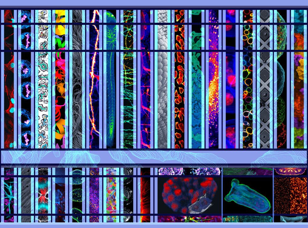
Neuronal connections
From new insights into visual processing to the nervous system of a marine worm, EMBL’s research in 2021 covered a wide range of neuroscience topics.
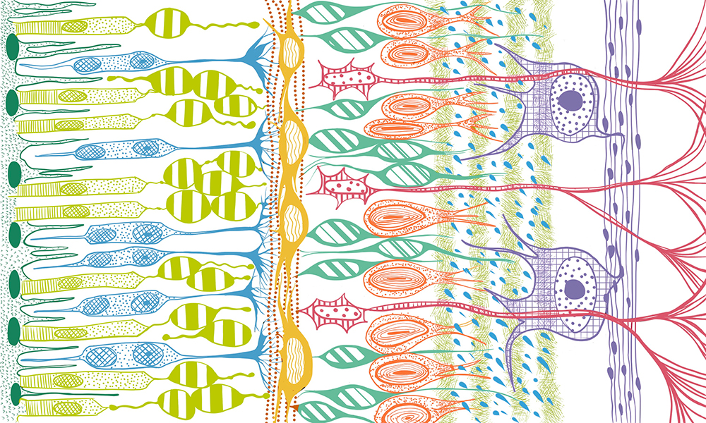
In 2021, scientists at EMBL created the most detailed dataset for an entire animal to date using electron microscopy and expression data. The cellular atlas of the marine worm, Platynereis dumerilii, will give an insight into the origin and evolution of the nervous system. The researchers showed that certain cellular structures known as mushroom bodies are a sensory organ on their own and that they express genes that are also activated in an important type of nerve cells in vertebrates.
A pioneering technique developed by researchers at EMBL allows neuroscientists to image live neurons deep inside the brain at high resolution. The technique is based on two state-of-the-art microscopy methods, three-photon microscopy and adaptive optics. It is a leap towards developing more advanced non-invasive techniques to study live tissues.
Based on a computational model of the retinal network, scientists at EMBL Rome were able to derive predictions about retinal physiology and test them experimentally. The study revealed an unexpected role for retinal cells in pre-processing visual information. These new insights could lead to the development of future prosthetic visual aids that mimic the retina’s visual processing.
Edit
“To make the [cellular atlas] data accessible to the community, we developed an open-source plugin. This has since turned out to be a very useful generic tool for multimodal big image data sharing and exploration.”
– Christian Tischer, Scientist / IT Engineer, EMBL Centre for Bioimage Analysis

“In a pandemic that once paralysed the world, EMBL found itself shouldering even more social responsibilities by doing what it can do best: making a difference through outstanding scientific contributions and discoveries.”
– Cristina Policarpi, postdoc, Hackett Group at EMBL Rome
Explore more 2021 EMBL neuroscience research highlights:
References
Vergara HM, Pape C, Meechan KI, Zinchenko V, Genoud C, Wanner AA et al. (2021). Whole-body integration of gene expression and single-cell morphology. Cell, 10 August 2021. DOI: 10.1016/j.cell.2021.07.017
Streich L et al. (2021). High-resolution structural and functional deep brain imaging using adaptive optics three-photon microscopy. Nature Methods, 30 September 2021. DOI: 10.1038/s41592-021-01257-6
lasiuk A, Asari H. (2021). Feedback from retinal ganglion cells to the inner retina. VPLoS One, 22 July 2021. DOI: 10.1371/journal.pone.0254611