Edit

Tag:

schaffitzel

Year
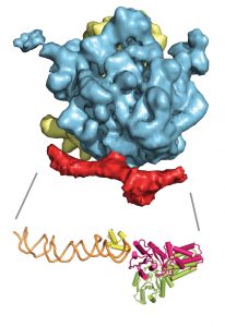
12 December 2010 This cryo-electron microscopy image shows the 3D structure of the ribosome (yellow/blue) bound to the signal recognition particle (SRP) and the SRP receptor (both in red). Below it is an atomic model of SRP (green-yellow/orange) and its receptor (pink). Image credits: EMBL/Schaffitzel.

How cells export and embed proteins in the membrane

Science Like an overprotective parent on the first day of school, a targeting factor sometimes needs a little push to let go of its cargo. Scientists at the European Molecular Biology Laboratory (EMBL) in Grenoble, France, have visualised one such hand-over. They were the first to determine the structure…

2010

science

No matching posts found

EMBLetc.

Looking for past print editions of EMBLetc.? Browse our archive, going back 20 years.

EMBLetc. archive

Newsletter archive

Read past editions of our e-newsletter

For press

Contact the Press Office
Edit