A no-brainer
Have you ever wondered what reflex testing is about? Why does your doctor tap the space below your knee with a hammer to see if your leg kicks forward? At the centre of this involuntary reaction is the muscle spindle, of which you can see a close-up in today’s Picture of the Week.
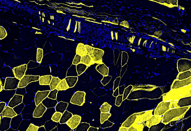
Muscle spindles are stretch receptors that are embedded inside skeletal muscles and primarily detect muscle contractions. Here, you can see the connection between neuronal fibres (yellow vertical stripes) and the muscle cells (nuclei stained in blue) inside the muscle spindle. The nerve cells transmit information about the stretch of the muscle to the spinal cord or brain.
When the knee reflex test is performed, information about the stretching of the muscles takes a shortcut to the spinal cord and does not enter the brain. From there it goes back to your thigh muscle to trigger the kick. This way, the reaction to the blow is very fast and tells the doctor that your nerves are working well. Since the brain is not involved, you would also not be able to stop the kick.
If you have a stunning picture of your science, your lab or your site, you can submit it here.