Swimming against the tide
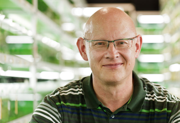
Give EMBL alumnus Jochen Wittbrodt ten minutes of your time, and he’ll convince you just how much biologists have to gain from embracing the impossible, pursuing outlandish ideas, and living on the edge
Wittbrodt clashed with the impossible in his very first position as group leader, at the Max Planck Institute for Biophysical Chemistry in Göttingen. He decided to run genetic screens on Medaka fish. In a genetic screen, scientists introduce mutations into an organism’s genes, and systematically look at the effects of those mutations on aspects like physical traits, physiology and behaviour. This is a laborious process – and was even more so back in the early 1990s, when gene sequencing was in its infancy – so the labs carrying out such studies tended to rely on a large workforce, and focus on established model organisms such as zebrafish. So Wittbrodt’s proposal to tackle a species for which no-one had yet developed specific tools, with a lab of 6 people, was met with scepticism. “But we just did it, and it worked,” he chuckles.
Birth of SPIM
A decade later, he had set up a lab at EMBL, and a presentation at a conference sparked another daring idea. “Pete Curry was showing an image of a whole fish in 3D imaged by a new method called OPT,” he recalls. “When I came back to Heidelberg, I met Ernst Stelzer in the EMBL cafeteria, and I said: ‘We need that, but we need it in live fish!’” Wittbrodt eventually convinced Stelzer to put a student on the job, and single-plane illumination microscopy (SPIM) was born. The basic idea of SPIM sounded like science fiction, says Wittbrodt: having a living organism under the microscope, shining a sheet of light on one layer of cells at a time, and putting that information back together to get a picture of the whole animal. The technique enabled Wittbrodt’s lab to see a fish embryo’s beating heart, and to trace its optic nerve from eye to brain. But he saw an opportunity to probe deeper – another ‘wild idea’: tracking each cell in an embryo, as they divide and move throughout the first day of its life. “Textbooks said you couldn’t do this in a vertebrate,” he says, “until Philipp Keller, who was then a PhD student in the Stelzer group, developed Digital Scanned Laser Light Sheet Microscopy (DSLM), and we saw how long we could image embryos for – and then we went for it!” The result, which they dubbed the Digital Embryo, earned them recognition from Science as one of the breakthroughs of the year for 2008.
If I have an aim, I’ll find a solution
Since leaving EMBL, Wittbrodt has continued to work on further developments to the technology, collaborating with Lars Hufnagel in particular. He has another wild idea up his sleeve. “You’d have the fish swimming freely, and passing through a laser scanner, being imaged as it goes.” Such a naturalistic setting would allow scientists to probe questions that are currently out of bounds, but so far, this particular idea has proven too ‘sci-fi’ for even Wittbrodt to get anyone to bite on.
How does he keep up his own drive in such situations, and keep swimming against the current? “Some people see the problems along the way, but forget what we’re aiming at. I think I can worry about a problem, but if I have an aim, I’ll find a solution, a way around it,” he replies.
Radical vision
Fostering and developing this type of unconventional approach is something that Wittbrodt has built into the workings of his lab at the University of Heidelberg. “I always give some ‘crazy vision’ talk once a year, at one of our lab meetings,” he says. One of the ideas he presented at this year’s talk was a new way to probe what conditions stem cells need in order to grow. His vision: to print an unconventional 3D scanner. “This scanner would not be a scanner in the sense that it scans your material; instead, your material – in our case stem cells – would scan the scanner,” he explains. The idea would be to use 3D printing technology to create a habitat for cells; hundreds of thousands of alcoves with different properties – some spongier, some harder, some with one type of food, others with another… Wittbrodt and colleagues would place their stem cells on this habitat, and let the cells choose their niche. “And we could just read out the properties and know what the stem cell needs,” he says, noting that he already has engineers, chemists and physicists, as well as specialists in 3D printing, on board.
Biologists have to be conversant in other fields in order to dream up – and implement – these approaches, he emphasises. “Specialisation gives you more details, and will keep some people happy,” he concedes. “But there are others who like to maybe bridge the boundaries and live in the interface, and try to get new ideas at the edges – this is much more the way I see myself. I wouldn’t say that I’m a zoologist, or a geneticist, or a cell biologist; I need a little bit of everything.”
To anyone who feels similarly, and has an ‘impossible’ idea that just might work, he says: “follow your heart; trust your gut!”