

Imagine: explore mitochondria
- Introduction
- A brief history of oxygen gas
- Journey of oxygen gas
- Mitochondria structure & function
- Cellular energy production inside mitochondria
- Image gallery
- Downloadable materials
Introduction
Mitochondria are membrane-enclosed organelles that produce the majority of ATP energy in many eukaryotic cells. Since their discovery in the 1850s, structure and function of these organelles have been heavily researched.
In this resource, we will explore how oxygen gas passes through the respiratory system and travels from the atmosphere into mitochondria organelles which use this gas to produce chemical energy in our bodies. We will also explore the membranous mitochondrial substructures in more detail using recent reconstruction images showing these organelles in their most natural state.
A brief history of oxygen gas
A brief history of the oxygen gas
With the evolution of oxygen gas generating cyanobacteria, atmospheric oxygen levels started increasing around 2.5 billion years ago. Back then, the majority of the planet’s inhabitants had adapted to a life without this gas. These organisms were collectively called anaerobic organisms. Such a drastic increase in oxygen levels wiped out many anaerobic species of the time. This event is also known as the oxygen catastrophe. An extinction event of that scale left a lot of room for oxygen-resistant organisms to flourish. Although oxygen gas was a deadly toxin to many life forms back then, today there are many organisms that cannot survive without it (including us, humans).
Currently, oxygen gas makes up for about 21% of our atmosphere and it is crucial to many life forms that generate adenosine triphosphate (ATP) using a chemical process called aerobic respiration. ATP is a type of cellular energy that cells use to perform vital tasks such as division and movement. In aerobic respiration, food particles such as glucose and fatty acids are consumed to generate ATP using the oxygen gas. In this resource, we will follow the oxygen gas and see where it travels inside our bodies to produce energy.
Oxygen gas in our planet is continuously replenished in a process called photosynthesis. Organisms such as the plants that you see around the EMBL campus produce food and chemical energy using the light rays from the sun and release oxygen gas as a byproduct. Let’s now follow how the oxygen gas travels through our bodies once it is released into the atmosphere.

Organisms breathe the oxygen gas in different ways
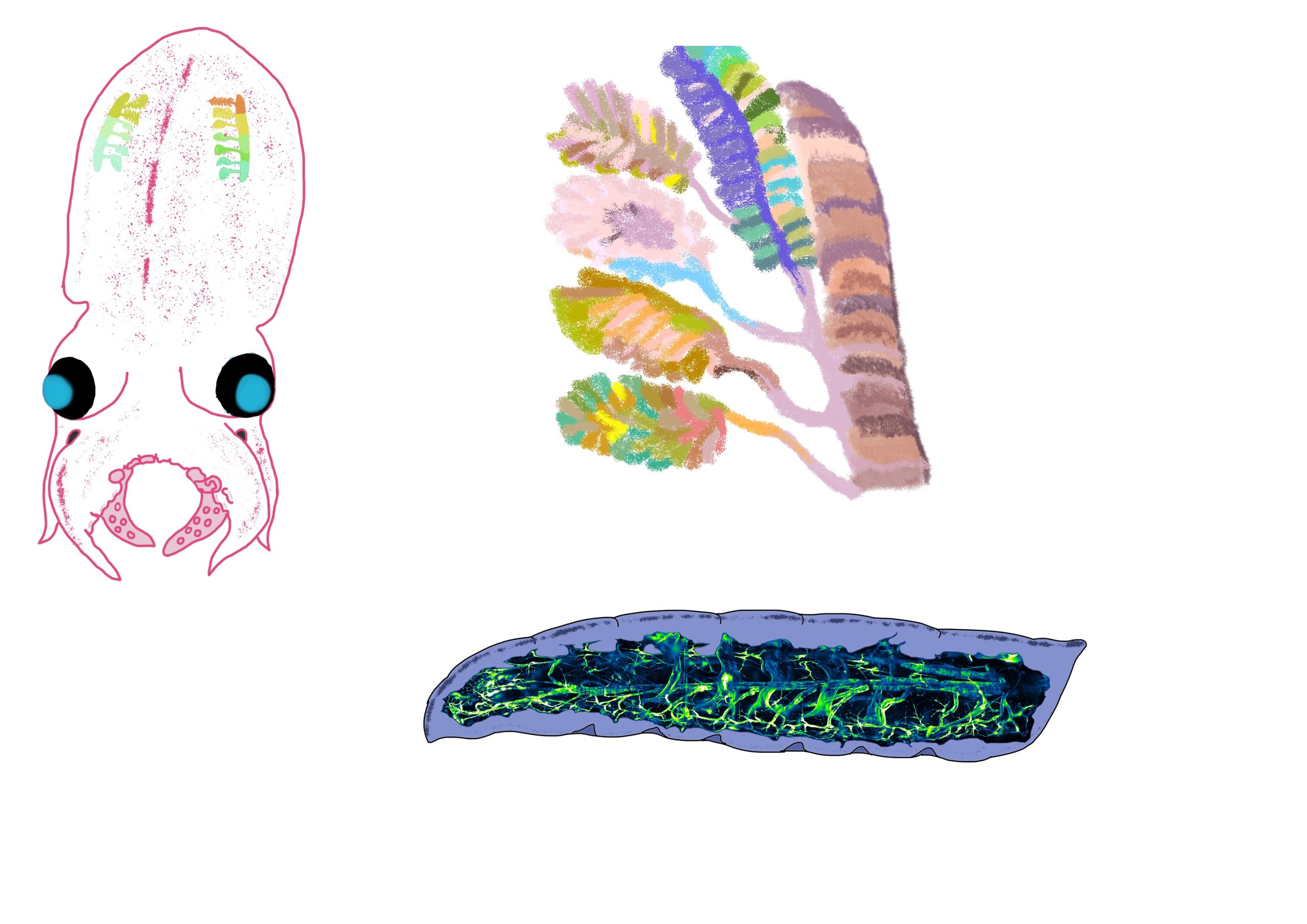
Organisms use different organs to breathe the oxygen gas. While mammals like humans use lungs, water dwelling animals such as the octopus use their gills to breathe in the water. Octopuses take in oxygen-rich water into their gills where the oxygen molecules diffuse into the circulatory system. Following this, used water is expelled from the gills. This expulsion of water also contributes to the locomotive abilities of these organisms.
Octopus gills are specialised filamentous organs that are reminiscent of trees. This branched and filamented structure has increased surface area which allows for more efficient exchange of oxygen and carbon dioxide gas per breath. Because these organisms and many other water inhabitants like fish, shrimp and mussels rely on the presence of enough oxygen gas in the water, oxygen rich clean waters are critically important for these organisms’ survival.

On land, organisms that breathe in the air from the atmosphere use structures like lungs and tracheal systems. While we are most familiar with lungs as respiratory organs, insect tracheal system is the most common type of breathing structures in land animals. Insect tracheal systems also have a branched like network for efficient oxygen gas exchange and this system runs along the insects’ body. Imagine having many many small nostrils coming out of your arms and legs that help you breathe… Insect use a similar strategy to breathe in the oxygen gas using their tracheal systems.
Journey of oxygen gas
From atmosphere to mitochondria
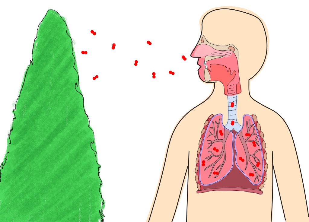
Oxygen molecules (in red) released from the trees such as the one you see below are taken into your lungs through breathing. Inside the lungs, it travels into the lung alveoli to enter the bloodstream.
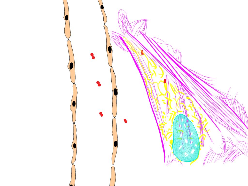
Lung alveoli or alveolar air spaces are composed of specialised cells with increased surface area to allow for more oxygen gas to be absorbed into the bloodstream. You can see an example of the alveolar air space shaped like a sphere below. Alveolar air spaces are very close to the blood vessels that connect with the rest of the body. This way, the oxygen we breathe enters the alveolar air space in our lungs and from there, it travels into the blood vessels to be carried to other parts of the body. Once inside the bloodstream, the oxygen gas is carried to individual cells of the body on haemoglobin proteins inside red blood cells (not shown).

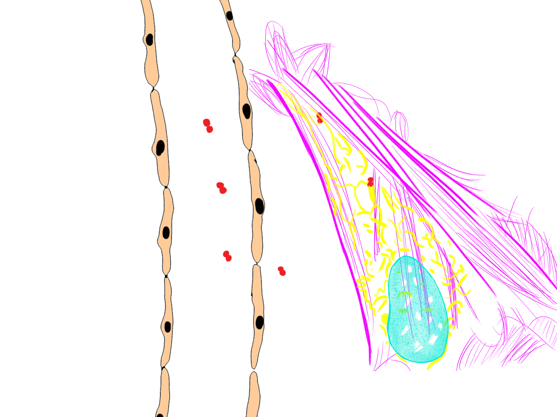
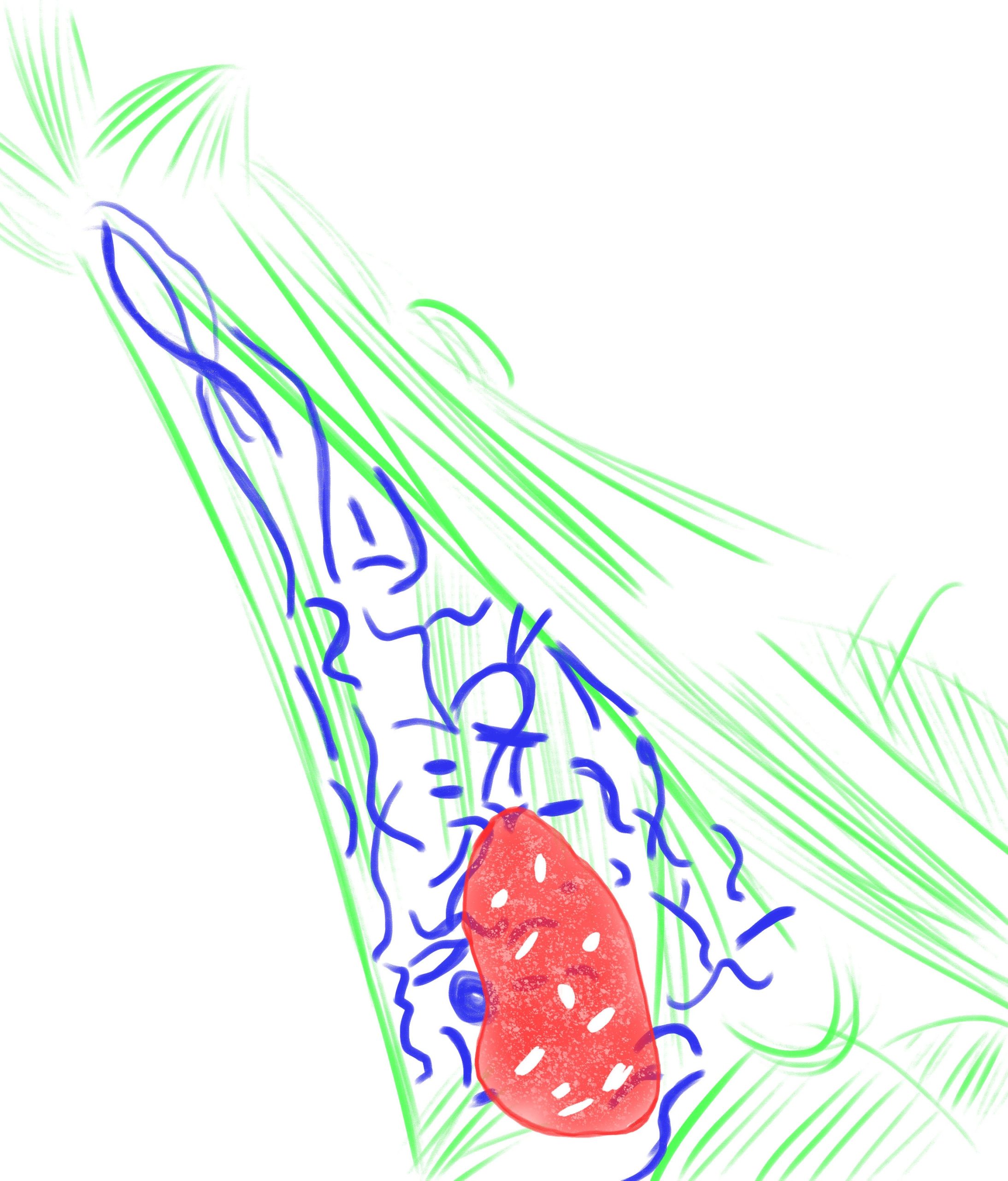
An example cell which is receiving the oxygen gas from the bloodstream is shown below. Oxygen gas travels through the cell membrane of the cell, entering the cytoplasm where it reaches the network of mitochondria (in yellow) where it is used in energy production. You can also see the cell nucleus stained in turquoise blue and the cytoskeleton in magenta.
Mitochondria structure & function
Mitochondria form bundles in cells
Mitochondria exist as intricate networks inside eukaryotic cells. A more zoomed in version of a part of this network is shown below. In case of a dysfunctioining mitochondrion in the network, such arrangement makes sure that other mitochondria can fuse with the dysfunctional one and help it complete the necessary functions.

Mitochondria have an extended network of inner membrane where energy production takes place.
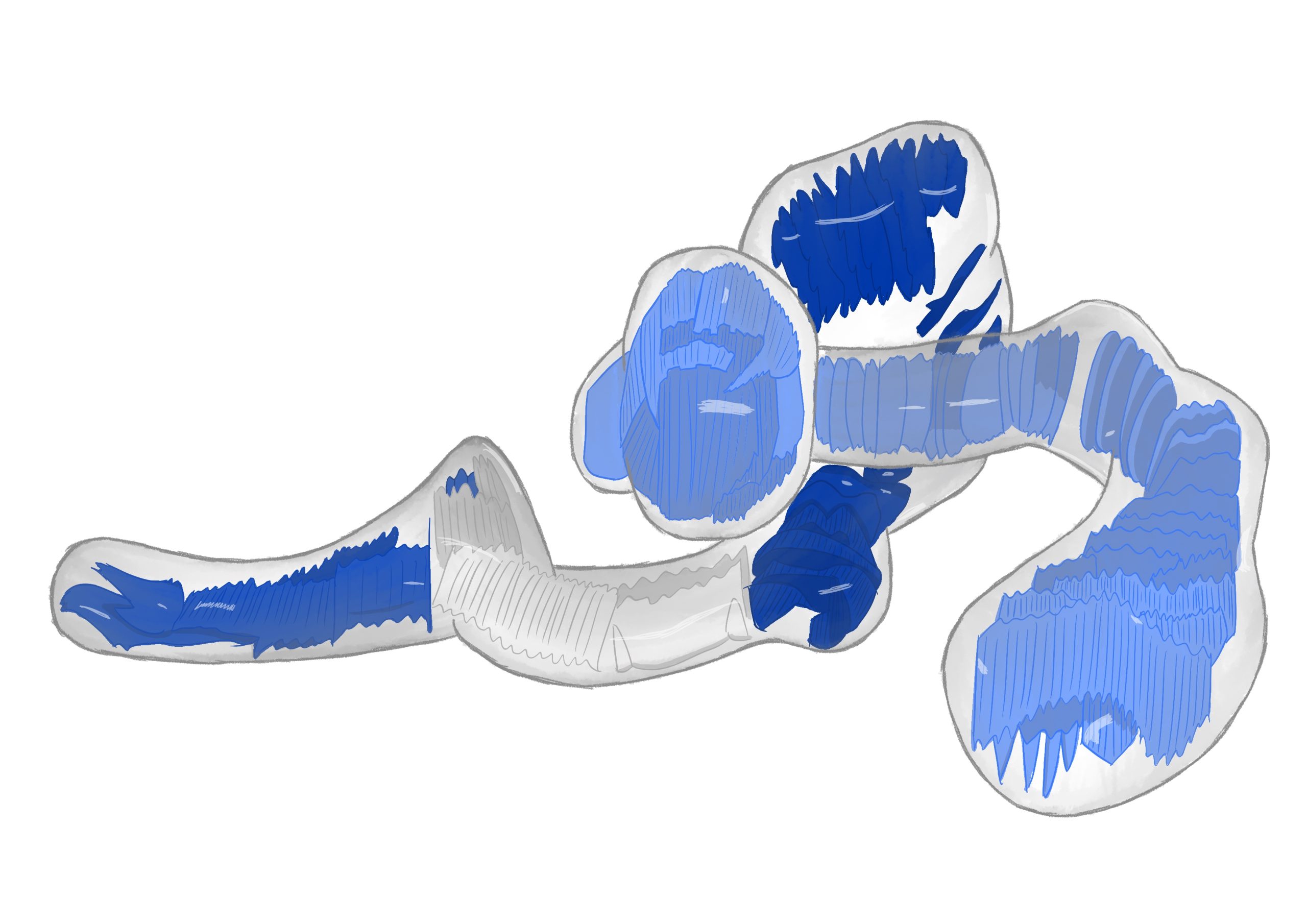
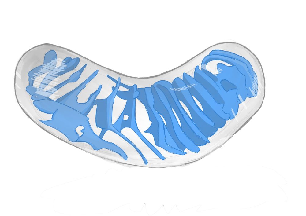
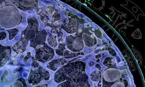
Below is an illustration of a single mitochondrion captured by EMBL scientists. The illustration shows the individual mitochondrion of human cells at their most natural form. As you can see, the mitochondrion has an extensive inner membrane network (in blue) that creates the space for cellular energy (ATP) production.

Cellular energy production inside mitochondria
Mitochondrial inner membrane is decorated by electron transport chain proteins
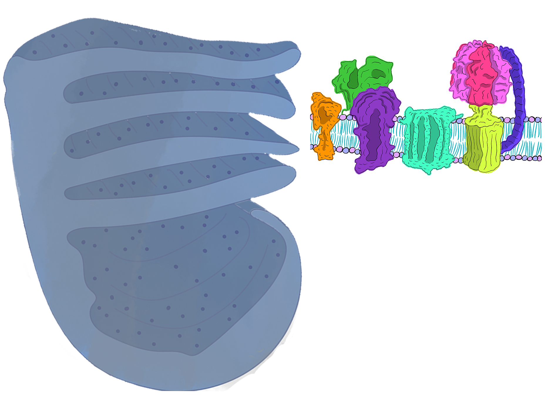
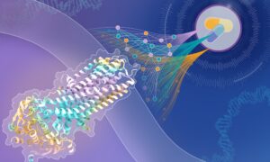
The mitochondrial inner membrane creates an extensive surface area for energy production. The inner membrane manueuvres inside the matrix and forms finger like protrusions. The reason that the inner membrane forms these finger like extensions is to create more available space for electron transport chain (ETC) to exist. Electron transport chain proteins decorate the mitochondrial inner membrane and function as sites of ATP production.

Cellular energy production takes place at the electron transport chain
Electron transport chain produces ATP by harvesting the chemical energy we collect from food such as glucose and fats. Chemical energy from the food we consume enter the mitochondria in the form of protons and energy rich electrons. As high energy electrons pass through the ETC, more proton ions are pumped into the inter-membrane space. Notice how protons are more concentrated at the inter-membrane space than in the matrix in the animation below. This concentration gradient is how mitochondria store energy much like electric batteries do. ATP Synthase enzyme then uses this stored energy to create ATP molecules from ADP. During the process, harvested protons come together with the oxygen gas and produce water, completing the process of cellular energy production. Because oxygen gas is used to complete the process of ATP production where a phosphate group is added to ADP molecules, this process is also known as oxidative phosphorylation. Watch the animation below to see how electrons move through the ETC complex and how ATP synthase produces ATP from ADP molecules.
Topic area: Cell biology
Age group: 14-16
Share:


Science Education and Public Engagement
Our inspiring educational experiences share the scientific discoveries of EMBL with young people aged 10-19 years and teachers in Europe and beyond. We belong to EMBL’s Science Education and Public Engagement office.