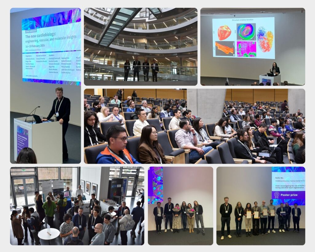
Best poster prizes at ‘The new cardiobiology: engineering, vascular, and molecular insights’
This year’s edition of the EMBL Conference ‘The new cardiobiology: engineering, vascular, and molecular insights‘ took place from 16 – 19 February 2026 at EMBL Heidelberg and virtually.
Speakers across cardiac and vascular biology, tissue engineering as well as systems biology came together to present new findings in the vascular biology and engineering to further strengthen the systems biology approach.
The focus was on developments in vascular engineering, cardiovascular regeneration, modelling the cardiovascular system in synthetic 3D multicellular systems, cardiometabolic mechanisms leading to both vascular and cardiac complications, the contribution of cardiovascular communication to multi-organ disease, and new translational breakthroughs.
224 people attended this conference on-site and 44 virtually. With a total of 128 posters to view, we held two poster sessions during which the presenters could discuss their research and their work was afterwards voted for by all participants.
There were seven poster prizes awarded during the meeting. We are pleased to share with you six of the winners’ abstracts!
Substrate stiffness induces senescence via the secretion of YBX1 by endothelial cells
Presenter: Colin Bodemer
Authors: Colin Bodemer, Büşra Nur Toğru, Ilka Wittig, David Rodriguez Morales, David John, Sandra Hemkemeyer, Maike Frye, Stefanie Dimmeler and Guillermo Luxán

Goethe University, Frankfurt, Germany
Abstract:
Cardiac ageing is marked by fibrosis, microvascular dysfunction, inflammation, and senescence-associated signalling. Fibrosis increases myocardial stiffness, but how endothelial cells respond to this age-related stiffening remains unclear. To address this question, we have cultured endothelial cells on stiffness matrices to mimic healthy and fibrotic myocardium using Sylgard 184, a non-reactive polymer. Endothelial cells cultured on stiffer matrices resembled aged cardiac endothelium. Specifically, they presented increased size, shorter telomeres, signs of senescence, increased gene expression of mesenchymalactivation markers and proinflammatory cytokines and reduced endothelial cell adhesion. Endothelial cells regulate their local microenvironment via the secretion of angiocrine signals. Treating naïve endothelial cells with supernatant of endothelial cells cultured on stiff matrices, impaired endothelial barrier, increased cell size, reduced nutrient uptake, and increased senescence.
Our data suggests that increased cardiac stiffening is an important trigger for the acquisition of ageing-related hallmarks in endothelial cells. Mass spectrometry of the supernatant from stiffness cultured endothelial cells identified several proteins enriched on stiff matrices, 30% of which were also upregulated in aged cardiac endothelial cells from snRNA-sequencing. Y-box binding protein 1 (YBX1), a stress-induced extracellular mitogen, was found enriched in stiff supernatant. Treatment of endothelial cells with YBX1 reduced nutrient uptake, increased expression of cell cycle inhibitors and led to an significant increase in β-galactosidase activity.
Our findings suggest that the stiffness induced secretion of YBX1 may induce endothelial cell senescence. Interestingly, spatial transcriptomics analysis of young and old murine hearts revealed that the expression of YBX1 is highly enriched in senescence hotspots in the heart. These findings highlight the importance of passive tissue stiffness and YBX1 during cardiac ageing and disease
Due to the confidentiality of the unpublished data, we cannot share the poster.
Cytomegalovirus latency exacerbates cardiac inflammation and tissue
remodeling after myocardial infarction
Presenter: Eleni Dapergola
Authors: Eleni Dapergola, Gustavo Campos Ramos, Marko Sustic, Georg Gasteiger, Diyaa Ashour, Peter Rainer, Roland Jahns, Stipan Jonjic, Laura Schreiber, Ulrich Hofmann,Ilija Brizic, Stefan Frantz, Emmanuel Saliba, Clement Cochain, Encarnita Mariotti-Ferrandiz

Würzburg Institute of Systems Immunology, Germany
Abstract:
Background: Epidemiological studies have consistently associated cytomegalovirus (CMV) seropositivity with adverse cardiovascular outcomes. However, the mechanisms by which CMV infection impacts pathophysiological mechanisms in the heart remain poorly understood. In this study, we sought to dissect how latent murine CMV infection impacts cardiac immune cell dynamics at steady-state and during post-myocardial infarction (MI) repair.
Methods: Experimental MI studies were conducted in C57BL/6J mice previously infected with murine CMV (MCMV). In situ inflammatory responses were characterized by spectral flow cytometry, bulk and single-cell RNA / T cell receptor sequencing, whereas cardiac function was monitored by echocardiography and magnetic resonance imaging (cMRI). Moreover, we retrospectively assessed the CMV serostatus in a well-characterized patient cohort with longitudinal cMRI data available and performed bulk T cell receptor sequencing on peripheral blood and myocardial samples to identify CMV-specific TCRs.
Results: Our findings show that exposure to MCMV induces long-term changes in the cardiac transcriptional profile and alterations in cardiac-resident immune cell populations, including the establishment of virus-specific memory CD8+ T cell residency. Compared to infarcted controls, mice previously exposed to MCMV exhibited stronger inflammatory response, marked by increased CD8+ T cell infiltration, and worsened cardiac function following MI. These observations in mice were supported by data from CMV-seropositive MI patients, who harbored CMV-responsive T cells in the heart.
Conclusions: Our findings demonstrate that latent CMV infection leads to long-term changes in the cardiac microenvironment, which ultimately impair post-MI healing outcomes.
Differential modulation of epicardial activation via the Fibulin-2–Nupr1b axis dictates cardiac regeneration after injury
Presenter: Gülsüm Kayman
Authors: Gülsüm Kayman, Gursimran Kaur, Shaoqiu Zhang, Séverine Leclerc, Emilie de Chantal, Darrell Belke, Gregor Andelfinger, Justin Deniset, Rubén Marin-Juez

University of Montreal CHU Ste-Justine Azrieli Research Center, Canada
Abstract:
In adult mammals, cardiac injury typically results in permanent fibrosis and impaired organ function. In contrast, the adult zebrafish heart exhibits a remarkable regenerative capacity, where the fibrotic response is a transient and tightly regulated phase that supports tissue regeneration. The mechanisms that prevent excessive fibrosis while promoting effective regeneration remain poorly understood.
Here, we identify the extracellular matrix protein Fibulin-2 (Fbln2) as a key regulator of epicardial activation and the differentiation of epicardial α-SMA+ myofibroblasts during cardiac regeneration. Using genetic tools to manipulate fbln2 expression levels, we show that attenuation and suppression of epicardial activation have opposite effects on long-term regeneration. Notably, despite reducing early cardiomyocyte proliferation and TGF-β-mediated epicardial activation, downregulation of fbln2 enhances regeneration by promoting faster collagen resolution and limiting fibrotic remodeling. Conversely, complete loss of fbln2 further suppresses epicardial activation and TGF-β signaling, impairing fibrosis resolution and leading to scar persistence.
Mechanistically, we identify epicardial Nupr1b, a stress-response factor, as a downstream effector of Fibulin-2 in controlling epicardial α-SMA+ myofibroblast differentiation. Using new gain- and loss-of-function tools, we found that Nupr1b mediates Fibulin-2–dependent epicardial responses following injury. We show that nupr1b mutants recapitulate fbln2 mutant phenotypes, whereas epicardial-specific overexpression of nupr1b restores cardiomyocyte proliferation and myofibroblast differentiation in fbln2 mutants.
Together, our findings identify Fibulin-2 as a key regulator of the balance between cardiac fibrosis and regeneration, mediated through TGF-β-dependent epicardial activation.
Moreover, our study uncovers a previously unrecognized Fibulin-2–Nupr1b signaling axis that directs epicardial myofibroblast differentiation. These findings provide new mechanistic insights into how modulation of epicardial cell state transitions through Fibulin-2–Nupr1b signaling regulates regenerative responses after cardiac injury.
Rare functional variants in FBN2 are strong genetic determinants for bicuspid aortic valve disease
Presenter: Laura Koebbe
Authors: Laura Koebbe, Katharina Knoll, Baravan Al-Kassou, Frank Oeffner, Fady Marcous,Sara Balzan, Dinara Sharipova, Jessica Bigge, Sandra Muerb, Sebastian Zimmer, Stefanie Heilmann-Heimbach, Maximilian Billmann, Marius Schwab, Martina Dreßen, Stefanie Doppler, Heribert Schunkert, Markus Krane, Paola Mass-Sanchez, Steffen Luetke, Philipp Barnowski, Franz-Georg Hanisch, Manuel Koch, Matti Adam, Victor Mauri, Malte Kelm, Tobias Zeus, Stephan Baldus, Wolfgang Pfuetzner, Markus Noethen, Teresa Trenkwalder, Carlo Maj, Salim Abdelilah-Seyfried, Gerhard Sengle, Johannes Schumacher

University of Bonn, Germany
Abstract:
Bicuspid aortic valve (BAV) is the most common congenital heart defect, defined by the presence of two instead of three valve leaflets. BAV is of great clinical relevance since it is a major precursor to serious cardiovascular complications. Given that its genetic basis remains incompletely understood, we aimed to identify novel risk genes and elucidate the underlying molecular mechanisms.
We conducted the largest whole-exome sequencing (WES) study of BAV to date, including 1,030 European cases. Gene-based burden analysis identified FBN2 as a novel genetic determinant, accounting for 2.1% of all cases. This represents a higher burden for BAV than the combined contribution of all previously established monogenic risk genes. Beyond BAV, carriers of FBN2 rare variants (RVs) frequently exhibited brachydactyly, suggesting a syndromic presentation involving cardiovascular and skeletal phenotypes.
Genetic validation using WES data from > 370,000 UK Biobank participants revealed an increased prevalence of aortic valve stenosis (AS) among FBN2 RV carriers, suggesting that they are also more frequently affected by BAV. Functional assays in patient-derived fibroblasts demonstrated that the RVs lead to FBN2 deficiency, promoting aberrant extracellular matrix remodeling and impairing the sequestration of transforming growth factor-β (TGF-β) and bone morphogenetic protein (BMP). Dysregulated TGF-β signaling during embryonic development appears to contribute to BAV formation, whereas enhanced BMP signaling in limb mesenchyme is implicated in brachydactyly. In vivo, fbn2b-deficient zebrafish show severe outflow tract valve malformations, confirming an essential role for fibrillin-2 in cardiac valve development.
In conclusion, FBN2 represents a novel and major genetic determinant of BAV.FBN2-related BAV defines a new fibrillinopathy characterized by combined cardiovascular and skeletal manifestations and should be particularly suspected in patients presenting with both BAV and brachydactyly. Future work will focus on integrating multi-omics data into a comprehensive human interactome to identify additional risk genes and cellular processes underlying BAV pathogenesis through network propagation and expansion algorithms.
Leveraging the human heart atlas to drive cardiomyocyte maturation in vitro
Presenter: Jack Palmer
Authors: Jack A. Palmer, Kazumasa Kanemaru, James Cranley, Emmanuel Wong, Nadav Yayon, Claudia Semprich, Rakesh Kapuge, Krzysztof Polanski, Andrew R. Bassett, Richard C. V. Tyser, Sarah A. Teichmann
Abstract:

Cambridge Stem Cell Institute, University of Cambridge, UK
Recent advances in single-cell and spatial omics technologies have significantly propelled our understanding of cellular diversity, function and interaction in in vivo cardiac tissue. Our lab has previously generated multimodal atlases of the adult and developing foetal heart, combining epigenetic, transcriptomic and spatial data to comprehensively profile cardiac cells. This has revealed a phenotypic spectrum of cardiomyocytes, ranging from the working myocardium of the ventricles to the components of the conduction system. This provides a cross-lifespan roadmap of the regulation underlying cardiomyocyte fate specification and maturation. Our regulatory roadmap is guiding our efforts to enhance cardiomyocyte maturation in vitro.
While in vitro cardiomyocyte models hold promise for regenerative medicine and disease modeling, they currently exhibit immature, foetal-like phenotypes. Using single-cell sequencing, we have profiled in vitro matured cardiomyocytes and integrated this data with our in vivo atlas to identify regulatory differences. We are actively targeting these differences through cytokine treatments and genetic perturbations to promote more adult-like maturation.
Our integrative approach enables systematic benchmarking of in vitro cardiomyocyte models against in vivo references, guiding the development of more physiologically relevant in vitro systems.
Due to the confidentiality of the unpublished data, we cannot share the poster.
Investigating the Role of Actin Regulators in Promoting CM Invasion During Zebrafish Heart Regeneration
Presenter: Bailin Wu
Authors: Bailin Wu, Florian Constanty, Lara Szilagyi, Arica Beisaw
Abstract:

Institute of Experimental Cardiology, Heidelberg University, Germany
Adult mammalian cardiomyocytes (CMs) are unable to proliferate robustly to replace lost myocardium following myocardial infarction (MI). MI ultimately leads to heart failure, which remains a leading cause of death in industrialized nations. In contrast, zebrafish harbor the ability to regenerate their heart, as CMs undergo dedifferentiation, proliferation, and ultimately replenish the fibrotic scar tissue at 60-90 days post-cryoinjury (dpci). We and others have shown that CMs extend protrusions into the injured area following apical resection and cryoinjury of zebrafish and neonatal mouse hearts, which is likely essential for cardiac regeneration and replenishment of the fibrotic scar.
To understand the underlying molecular and cellular mechanisms regulating invasion of newly proliferated CMs at the wound border and subsequent replenishment of scar tissue by functional CMs, we compared the transcriptomes of remote CMs and border-zone CMs by way of single-cell RNA-sequencing.We discovered new gene candidates that potentially regulate CM invasion during zebrafish heart regeneration. Among the pathways enriched in border zone CMs, we find that cytoskeleton organization is significantly upregulated. We find that actin cytoskeleton regulators are positively regulated by AP-1 transcription factors, which are essential regulators of the CM regeneration process. Using a newly generated transgenic model, we find CM-specific overexpression of a cytoskeleton regulatorleads to stabilization of F-actin in CM, promotes CM dedifferentiation, and accelerates scar resolution. Currently, more studies are ongoing to understand whether and how actin cytoskeleton organization participates in CM invasion.
Altogether, our data provide an in-depth characterization of a largely understudied process during cardiac regeneration in zebrafish. The results could benefit the development of cell-based therapeutic strategies to replace fibrotic scar tissue with functional CMs to increase cardiac function after MI.
Due to the confidentiality of the unpublished data, we cannot share the poster.
The EMBL Conference ‘The new cardiobiology: engineering, vascular, and molecular insights took place from 16 – 19 February 2026 at EMBL Heidelberg and virtually.