Development of cryo-electron microscopy at EMBL

Cryo-electron microscopy (cryo-EM) is a technique that determines the shape of frozen samples by firing electrons at them and recording the resulting images.

Cryo-EM is a relative newcomer to structural biology – the study of the structure and function of proteins and other molecular assemblies to understand how they form, work, and interact; how they malfunction in disease; and how to target them with drugs.

Originally, electron microscopy of biological material was hampered by a range of challenges, including a lack of a suitable preparation technique: a method that would allow researchers to prepare biological material for very precise measurements under the microscope whilst keeping it from being damaged.

SARS CoV-2 virus artistic render
SARS-CoV-2’s ability to infect cells relies on interactions between the viral spike protein (magenta) and the protein ACE2 (blue), which is present on the surface of human cells. These interactions can be disrupted by sybodies (black) – synthetic mini-antibodies similar to those produced by camels and llamas. Credit: Rayne Zaayman-Gallant/EMBL

Development of a revolutionary technique for structural biology

Working at EMBL in Heidelberg in the early 1980s, Group Leader Jacques Dubochet and technician Alasdair McDowall discovered that flash-freezing proteins in liquid ethane could hold them still and preserve their structure, a process called vitrification. This was a critical advance that laid the groundwork for the rise of cryo-EM and was a revolution in structural biology. In recognition of the importance of this work, Dubochet was one of three scientists awarded the 2017 Nobel Prize in Chemistry. EMBL provided Dubochet with a working environment that he described as unique in allowing him to "explore avenues which had not been explored". He expressed his gratitude to EMBL by donating an official replica of his Nobel medal to the organisation.

Today, cryo-EM is a game-changing technique for structural biology, enabling molecules to be imaged quickly and in the variety of shapes that they naturally adopt in the cellular environment. In recent years, the number of protein structures determined by cryo-EM has exploded; some experts believe that within the next five years, cryo-EM will become the dominant method for determining protein structures.

EMBL continues to play an important role in this field. Dubochet highlighted that EMBL not only enables researchers to access cryo-EM experimental services but also offers the necessary expertise to fully apply the technique to their research questions: "These methods are complicated and difficult and need to be used and exploited by many researchers. But the capability of exploiting them is by those who are at the cutting edge of cryo-EM." Cryo-EM clearly has come a long way since Dubochet and McDowall's crucial discovery at EMBL -- but as McDowall noted, "Everybody has to vitrify [prepare] the sample. That's still step one."

Jacques Dubochet holding his Nobel medal during Nobel week
Jacques Dubochet holding his Nobel medal during Nobel week. Credit: Alexander Mahmoud/Nobel Media

I am pleased to offer this copy of my Nobel medal to EMBL in testimony of my great thankfulness to an institution that, in my view, would deserve to be the laureate of the Prize.

Advancing scientific discoveries towards application

One area in which cryo-EM has led to a step change in the level and speed of progress is in the design and development of vaccines. Starting in the late 1990s, scientists from around the world used cryo-EM to determine the structures of viruses such as the hepatitis B virus, hepatitis C virus, Zika virus, rhinovirus C, dengue virus, and tick-borne encephalitis virus. EMBL researchers have played an important part, e.g. by providing insights into the structure of HIV.

Research on viral structures also formed part of Ilaria Ferlenghi's work at EMBL in the late 1990s -- experience she was subsequently able to bring to industry. Some 15 years after leaving EMBL, Ferlenghi -- now Head of Structural Vaccinology at GSK -- has built the company's cryo-EM programme for imaging proteins on the surface of viruses to identify candidate targets for new vaccines. This was possible because the technology has matured to the point where it can yield precise structural information suitable for drug development. Pharmaceutical companies around the world are exploiting cryo-EM for the development of new drugs and treatments for diseases.

As the COVID-19 pandemic unfolded, cryo-EM yielded precise images at an unprecedented pace. Only three weeks after Chinese scientists reported the first genome sequence of the virus, researchers reported the structure of the SARS-CoV-2 spike protein; a further five days later, they published images of the spike protein bound to its human target, angiotensin-converting enzyme 2 (ACE2). These insights provided important information that was used in the development of COVID-19 vaccines.


Next revolution of cryo-EM

EMBL continues to push methodology development further. The group of John Briggs at EMBL pioneered cryo-electron tomography (cryo-ET), a technique with origins in cryo-EM. While cryo-EM examines highly purified molecules to determine their shapes, cryo-ET allows scientists to take 3D snapshots of molecular interactions inside the cell. 
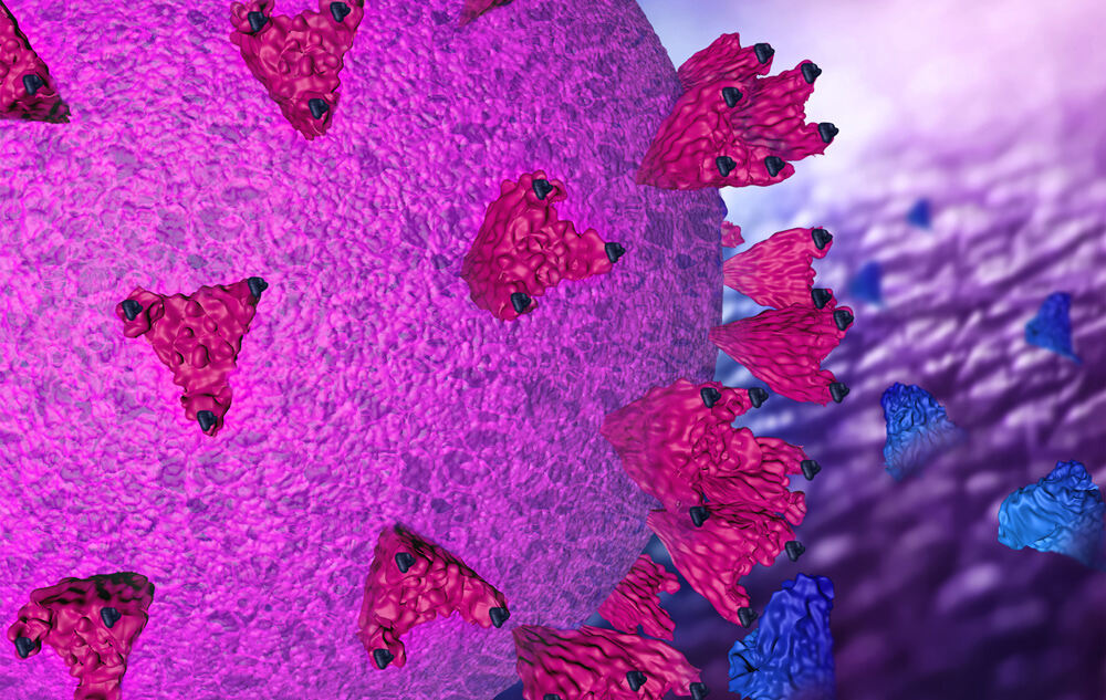
This is important, because many complexes cannot be purified; information on both the structure and the location of large molecular complexes in the cell is crucial for understanding their function. Now at the Max Planck Institute of Biochemistry in Munich, Germany, Briggs was the first to achieve extremely precise visualisations of a biological structure using cryo-ET -- areas of the protein shell of HIV.

Recently, a group at EMBL led by Julia Mahamid used the same techniques to observe a key molecular machine in action: bacterial RNA polymerase, which turns the organism's genetic code into RNA and protein. Her work has been described by other researchers as "a technical breakthrough". Mahamid highlighted the importance of EMBL in achieving these advances: "We couldn't have produced this quality of research within just three years of starting a lab without the data and support we have at EMBL. This isn't an incremental advance, but rather a jump. Cryo-EM revolutionised structural biology a few years ago. Our new approach [cryo-ET] is likely to contribute to a second revolution where you can study structures directly while [they are] still inside cells".

EMBL continues to develop imaging methods in collaboration with external research groups. "We [EMBL] help to develop methods that are relevant to questions of researchers in external universities by collaborating with them to develop cutting-edge technologies before they become accessible as a service," said Mahamid. Recognising the facility's exceptional instrumentation, its willingness to fine-tune hardware and software as needed, and the lack of bureaucracy in accessing the service, Albert Weixlbaumer from the Institute of Genetics and Molecular and Cellular Biology (IGBMC) in Strasbourg, France, commented: "The experience at EMBL was as good as it can get. [...] The quality and amount of data we obtained is difficult to match at any of the sites I know."

In 2020, Weixlbaumer's collaboration with EMBL resulted in a study revealing insights into bacterial gene expression that was published in the top-tier journal Science.


What the future holds

EMBL Heidelberg now hosts its state-of-the-art cryo-EM service in a new facility -- the EMBL Imaging Centre -- which offers a synergistic portfolio of imaging techniques to enable academic and industry users from the international research community to conduct new ground-breaking research. As Ferlenghi described:

"When you have many different techniques in the same place that can be combined, and you can discuss them with the different experts in each field, this is the advantage of having a place like EMBL. On top of that, the passion and curiosity that EMBL [scientists] have is different from what you can get from any other institution."

EMBL’s experimental services deliver substantial value to Europe by delivering scientific services and thereby enabling researchers to conduct novel and demanding experiments that could not be easily achieved at a purely national level, an independent review found.

The global consultancy Technopolis Group, specialising in research and innovation policy, conducted a survey and analysis of external (non-EMBL) users of EMBL’s experimental services. The survey aimed to identify whether users perceived scientific, technological, societal and economic benefits as a result of utilising the experimental services at EMBL Barcelona, Grenoble, Hamburg, Heidelberg, and Rome.