On the Workbench
A project to automate the preparation of Cryo-EM SPA grids with multiple samples on a grid and time resolved freezing. The production of associated nanometer scale 2D ice thickness maps will provide microscope direct access to potentially usable sample areas.
In consultation with electron microscopists of the EMBL Grenoble and Heidelberg sites.
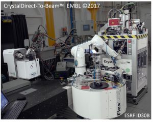
A project to integrate the CrystalDirect-to-beam concept at MASSIF 1, the future ESRF-EBS fully automated MX beamline ID30A1. Crystals supplied in CrystalDirect plates will be automatically harvested and data collected at cryogenic or at room temperature without manual intervention.
In collaboration with the Synchrotron Crystallography Team (M. Bowler, A. McCarthy), the High Throughput Crystallization team (J.A. Marquez) and the ESRF MX Group (G. Leonard)
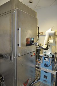
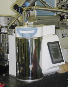
Designed to operate in a normal room, its built in temperature control system allows harvesting crystals at 4 and 20 deg Celsius. Two times faster than the previous generation, it offer 30 seconds harvest turnover time.
Download the CDh3 Harvesting video
In collaboration with the High Throughput Crystallization team (J.A. Marquez)
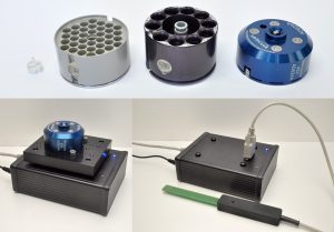
An electronic identification system based on Radio Frequency IDentification (RFID) tags compatible with cryogenic temperature (CryoRFID) is being developed to track samples stored in cryo containers, like MX pucks, Cryo-EM pucks or grid boxes. Specific readers are used to read the UIDs of the CryoRFID tags and to store/retrieve user information in the tags.
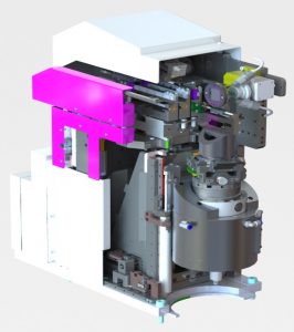
Optimized for serial data collection on solid supports and kinetic studies, it will equip the future ESRF-EBS micro-focus beamline ID29. A fast SSX scan head (up to 100 mm/s) and a synchronization system will allow shooting up to 2000 micro-crystals per second with x-ray photon bunches delivered by the beam chopper of the beamline.
Download the MD3upSSX scan video (50 mm/s line scans)
In collaboration with the ESRF MX group (D.de Sanctis) and the company ARINAX.
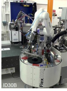
The ESRF-EBS MX beamline ID30B will see its FlexHCD sample changer equipped with miniSPINE single and double grippers, making it first miniSPINE pilot beamline opened to users.
Download the miniSPINE video
ID30B – a versatile beamline for macromolecular crystallography experiments at the ESRF.
The NewPin project
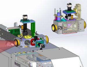
Two new BioSAXS sample exposure units are developed for the ESRF-EBS beamline ID29 and the EMBL@PETRA-III P13 beamline. The fist, to optimally expose sample solutions in thin glass capillaries, will offer in-situ absorption spectrophotometry and DLS particle size measurement to control the quality of the samples. More flexible, the second is a vacuum chamber with motorized stage and on beam axis camera. It is designed to expose sample solutions in micro-fluidic devices.
In collaboration with EMBL Hamburg (D. Svergun, C. Blanchet) and the ESRF (P. Pernot, M. Tully)
Macromolecular Crystallography
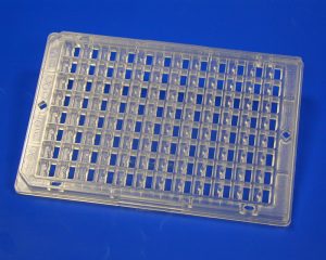
Designed to handle sample holders used in Macromolecular X-ray crystallography, the Flex robotics family is built around a 6 axis industrial robot. The family currently includes 3 members equipped with different storage Dewars. They are all provided with a tool changer and various handling tools.
FlexED8 (picture), a sample changer compatible with SPINE, NewPin and miniSpine sample holders that can hold up to 252 sample holders in a small footprint open Dewar with built in ice filtering system. Different cryo-grippers are used to safely transfer the sample holders to a goniometer. The sample exchange time can be as low as 4 seconds when the Twin SPINE cryo-gripper is used. FlexED8 was developed in the context of the NewPin project and tested on the EMBL/ESRF/INDIA BM14 CRG beamline.
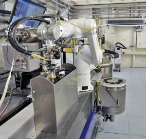
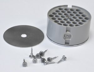
FlexHCD, a sample changer compatible with SPINE sample holders. It includes a modified High Capacity Dewar initially developed within the ESRF MX group. It can hold up to 384 samples in uni-pucks or 240 samples with vials in SC3 pucks, or a mix of the two. The sample holders in uni-pucks are transferred using a single or a Twin cryo-gripper and with the IRELEC-Alcen flipping gripper when stored with vials in SC3 pucks. FlexHCD is installed at the ESRF ID30B beamline and being duplicated to equip four other ESRF MX beamlines.
FlexED3, a cryo-storage unit associated to a CrystalDirect™ (see below) harvester to automatically store crystals harvested with SPINE sample holders in SC3 pucks. FlexED3 is currently in operation at the Grenoble HTX lab.
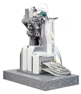
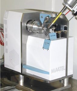
The CrystalDirect harvesting station
Operated by the HTX lab since 2017, it allows to automatically harvest, vitrify and mount on SPINE sample holders crystals of any size, as well as micro-crystals harvested in batches.With a storage capacity of several hundred samples in SC3 pucks or in uni-pucks and a turnover time of 1’30”, it is extensively used in fragment screening applications.
Inside look
From a collaboration with the High throughput crystallization team (J.A. Marquez)
TOP CrystalDirect™
Jointly with the EMBL Grenoble HTX Team, J A Marquez.
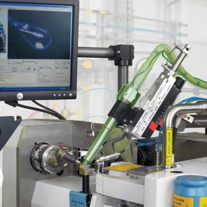
Automated crystal harvesting and in-plate crystal screening.
Download the ‘CrystalDirect Laser Harvesting’ video [9.6MB]
Download the ‘In-Situ data collection’ video [12.4MB] – EMBL/ESRF BM14 beamline, Hassan Belrhali
Tentative for a new European sample holder standard for cryo-crystallography
The NewPin project
https://www.moleculardimensions.com/products/alternative-crystal-mounting-systems
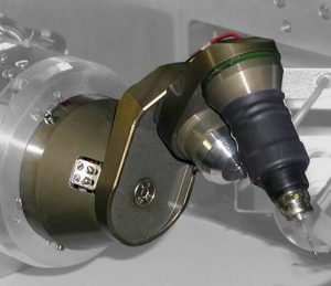
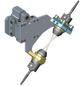
Towards a compact and precise sample holder for macromolecular crystallography.
In collaboration with EMBL@PETRA-III MX2, G Bourenkov and ARINAX.
Download ‘MD3 SOC-1’ video [508KB] – Kappa = 0°, Omega 160°/s; Glass needle 0.3 µm tip; Scale 1 µm/div
Download ‘MD3 SOC-2’ video [8.1MB] – Kappa = 48°, Omega 45°/sec; Glass needle 1 µm hole; Scale 10 µm/div
Download ‘MD3 4D scan’ video [4.1MB] – Needle Ø8 µm X 150 µm; 4D scan: 180°, 18°/s; Scale 10 µm/div
Download ‘HC1b – Humidity Controller for MX data collection experiments’ details [119KB]
Collaborations: Crystal Dehydration
ESRF, Diamond, MAX-Lab, Bessy, CLS
Download ‘Load/unload one sample (real-time)’ video [3.5MB]
ESRF ID14-1, ID14-2, ID14-3, ID14-4, ID29, ID23-1, ID23-2; embl-France ESRF BM14
Download ‘MK2 – mini Kappa goniometer head’ details [225KB]
Download ‘MK2 – mini Kappa goniometer head’ video 1 [3.4MB] – stability when mounted on a MD2 diffractometer @ Chi = 0, Phi fixed, Omega 90 deg/s. Grid 10 µm/div; vertical arrow 4 µm
Download ‘MK2 – mini Kappa goniometer head’ video 2 [5.2MB] – stability when mounted on a MD2 diffractometer @ Chi = 48 deg, Phi fixed, Omega 20 deg/s … 90 deg/s. Grid 10 µm/div; horizontal arrow 5 µm
ESRF ID14-4, ID29, ID23-1; embl-France ESRF BM14 CRG, ESRF BM30 CEA-IBS CRG, APS LS CAT; Diamond; ALS; Bessy
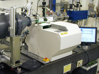
Download ‘MD2M – MicroDiffractometer’ details [303KB]
ESRF ID14-1, ID14-2, ID14-4, ID29, ID23-1, ID23-2
Download ‘MicroSpecrophotometer lens setup’ details [897KB]
Use it at the ESRF
ESRF ID14-x, DLS
Download ‘Microdiffractometer MD2’ details [1.7MB]
SLS X06S; ESRF ID14-3; embl-France ESRF BM14 CRG; APS NE CAT, LS CAT; Bessy, ALS, CLS, SSRL, EMBL-HH, Alba, MAX-lab, Soleil
Automatic Crystal Alignment Library C3D
Read more about C3D – a tool for automated crystal alignment on high throughput macromolecular X-ray beamlines – on the EMBLEM Technology transfer website.
ESRF ID14-1, ID14-2, ID14-3, ID14-4, ID29, ID23-1, ID23-2; EMBL/ESRF BM14 (CRG); NSLS
Download ‘OAV – On beam Axis Video-microscope’ details [356KB]
ESRF BM30 FIP CNRS/IBS GRG + EMBL/ESRF BM14 (CRG); NSLS

Download ‘1″ exposure, 5 passes’ video [336KB] – real-time 5 x 0.2″
ESRF ID14-1, ID14-2, ID14-3, ID14-4, ID29, ID23-1, ID23-2; APS NE-CAT, EMBL-HH
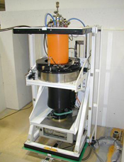
Neutron Diffractometers
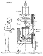
FINDer (Fast Imageplate Neutron Diffractometer)
FINDer is a Neutron Laue diffractometer using image plates (IP) as detector.
FINDer is the industrial version of the LADI quasi laue diffractometer that has been developed at the EMBL Grenoble outstation for protein crystallography (routinely used at the ILL since 1998). FINDer has an extended application field. It can be used either for material science (thermal neutrons, large He cryostat) or for biology experiment (cold neutrons).
The first unit has been constructed by the SICN company and is now installed at the ILL on the VIVALDI station. It sits at the end of a thermal neutron guide that provides a white beam (0.8<L<4.5 Å) that can be filtered to 0.8<L<3 Å or 3<L<4.5 Å.
The sample environment includes an ILL ‘Orange’ cryostat for1.5 K to 600 K with optional standard dilution inserts (down to 50 mK) and high-pressure cells up to 49 mm in diameter.
A time of flight analyser has been integrated in the diffractometer to monitor the tuning of the neutron band pass filter, and to record the neutrons energy spectrum together with the diffraction patterns.
Technical and commercial information:
EMBL: gpapp@embl.fr
EMBLEM
Principle
A neutron image plate is a storage phosphor plate sensitive to neutrons. Image plates are two-dimensional detectors. When exposed to neutrons, ‘colour centres’ are created inside the phosphor in quantity proportional to the neutron dose. As in photographic films, these colour centres makes a latent image. A red laser spot that releases the colour centre reads this latent image. Blue light is then emitted in a quantity proportional to the number of colour centres. While the plate is scanned, the intensity of the blue light is recorded as a function of the laser spot position, making then an intensity map of the latent image. Unlike photographic film, the plates are reusable after erasure by exposure to light.
The detector is a vertical drum internally covered with neutron image plates. The sample sits in the centre of the drum and can be rotated around the drum axis. Different sample holders can be installed, including cryo cooling systems. The diffractometer is designed to accept further extensions for magnetic sample environment. To hit the sample, the neutron beam passes through two holes in the drum.
The detector works in three phases: firstly, an intense light erases the image plates; secondly, the sample is exposed to neutrons and diffraction is recorded on the image plate; thirdly, a reading head scans the drum (the drum rotates while the head moves along it).
The reading head is composed of a PMT placed in front of the image plate. An optical system focuses a red laser diode on the image plate, just in front of the PMT, this excites blue photostimulable luminescence that is measured in the PMT. A filter in front of the PMT eliminates the red light. The PMT current is integrated on the surface of each pixel. The drum is scanned in ‘phonographic’ mode (a one pixel width spiral). The pixel values are stored on a computer disk and can be displayed as colour map images on a computer screen. A set of patterns can be collected automatically from one sample. For each acquisition, the sample parameters can be set (sample orientation, temperature…)
Main features
FINDER is a turnkey diffractometer based on an open architecture and modular design for flexibility and easy integration in the user environment.
- Compact design: 2x1x1 m (HxWxD), Mounted on 3 Air Pads
- Beam height: from 1100 to 1600 mm (other upon request)
- Compatible with different sample holders, including large He cryo cooling devices
- High performance image plate detector:
- Sensitive area: 800 x 400 mm (4/5 of the internal surf. of a Ø 320 mm, H 400 mm cylinder)
- Pixel Size: from 100 x 100 to 400 x 400 µm
- Readout time: 2 to 7 minutes, (400 to 100 µm pixels)
- Dynamic range: 16 bits
- Spatial resolution: 200 µm FWHM
- DQE: 40 to 60%
- PC / Windows NT based architecture
- PC/Windows NT-based architecture
- Ethernet interface for data transfer
- User friendly control software:
- Full control of the instrument through a GUI
- One access level for tests and tuning, one access level for experiment control
- Windows NT standard application written in Visual Basic (VB)
- Object oriented software using ActiveX components
- Ability to add experiment dependant controls (*temperature)
- Time Of Flight* (TOF) option for beam energy spectrum control
- Experiment control process can be modified easily (Visual Basic module)
Options
- Beam energy spectrum monitor (Time of Flight)
- Sample cryo cooling control