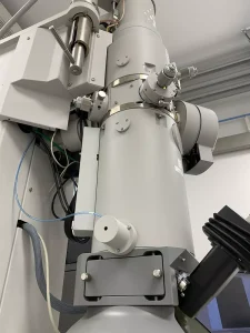
Tecnai G2 Spirit BT (T12)
120 kV accelerating voltage, LaB6 source. 16M CETA camera, used mainly for negative staining samples, but also capable of cryo-EM using side entry cryo-holders.
Since 2016, the Electron Microscopy (EM) facility provides advanced expertise in electron cryomicroscopy, from sample preparation to image analysis, with a focus on single particle analysis.
The equipment is maintained and managed by the facility staff, who also provide training and assistance for anyone wishing to use any of the equipment listed below.
Cryo-EM facilities are available for internal users and on a collaborative basis. Please get in touch with Sarah Schneider to discuss possible access.
120 kV accelerating voltage, LaB6 source. 16M CETA camera, used mainly for negative staining samples, but also capable of cryo-EM using side entry cryo-holders.
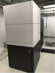
200 kV accelerating voltage, field emission gun (X-FEG) source. This microscope is equipped with an autoloader, Falcon 4i direct electron detector and SelectrisX energy filter. The setup is connected to WARP on-the-fly data processing node. This microscope is used for cryo-EM sample optimisation and data collection.
Supporting equipment:
Fischione 1070 PLasma Cleaner
Vitrobot Mark IV
PELCO EasiGlow glow discharger
Edwards carbon evaporator
We use 2PB storage system running on NetApp technology for cryo-EM data storage and processing. Additionally, a 200 TB parallel file system using BeeGFS technology is used as a scratch space for fast data processing.
The computing cluster shared with the Institut de Biologie Structurale (IBS) allows efficient data processing using state-of-the-art data processing software such as Relion or CryoSPARC. Eight cluster nodes comprising a total of 40 GPUs allow efficient data processing by multiple users. CPU cluster with 600 CPU provides additional resources for other software packages. Several dedicated GPU-servers are available for specialised usage (i.e. deep learning applications and on-the-fly data processing). High-performance computing at EMBL is supported by SBGrid.
Cryo-Electron Microscopy Specialist
ORCID: 0000-0001-5199-5157
Edit
Group Leader
wgalej [at] embl.fr
ORCID: 0000-0001-8859-5229
EditCryo-electron Microscopy Expert
ORCID: 0000-0001-9226-3382
EditEMBL Grenoble also participates, together with the other Partnership for Structural Biology (PSB) institutes, in running the CM01 Titan Krios cryo-electron microscope at the ESRF for external users.
For more information regarding access, you can visit the ESRF page or contact EMBL staff scientist Michael Hons.