Correlative light and electron microscopy (CLEM) is a collection of methods that allows us to locate a molecule of interest within a high-resolution cellular map.
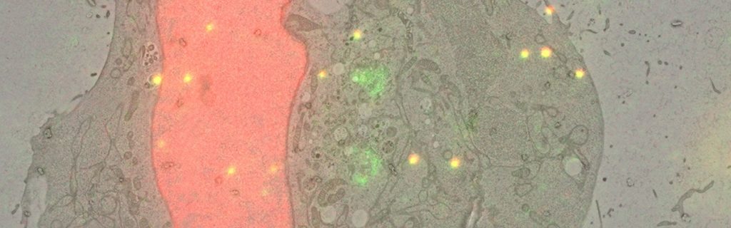
This e-learning video focuses on a specific method where we preserve the fluorescence throughout the electron microscopy (EM) sample preparation. This allows us to image the same section with the light and electron microscope, so that we can overlay the information coming from both imaging modalities with very high precision.
Target audience
This video is targeted at scientists interested in subcellular localisation of a protein of interest by combining light and electron microscopy.
This video takes you step-by-step through the method. However, because of its complexity, a basic level of knowledge in sample preparation for EM and ultramicrotomy would be advantageous.
Learning objectives
• Prepare sample used for on-section CLEM
• Evaluate tricks for light and electron microscopy imaging
• Use methods to obtain high precision correlation of the light and electron microscopy images using special beads/fiducials and dedicated software solutions
CLEM protocols
• Precise, correlated fluorescence microscopy and electron tomography of lowicryl sections using fluorescent fiducial markers. Kukulski W, et al. (2012). Methods Cell Biol. 111. 235-57. Europe PMC link
• eC-CLEM: A multidimension, multimodel software to correlate intermodal images with a focus on light and electron microscopy. Heiligenstein X, et al. (2017) Methods Cell Biol. 140. 335-52. Europe PMC link
Feedback
What did you think? We hope you enjoyed this e-learning video and we’d welcome your feedback and suggestions for future courses.
Project team
Project manager: Jacqueline Dreyer-Lamm
Executive producer, scientific advisor and voice over: Rachel Mellwig
Technical guidance: Pedro Machado & Paolo Ronchi
Editor and scientific oversight: Yannick Schwab
Video producer and motion graphics: Claudiu Grozea